. 2016 Aug 15;8(8):3471-81.
eCollection 2016.

Aβ mediates Sigma receptor degradation via CaN/NFAT pathway

Affiliations

Aβ mediates Sigma receptor degradation via CaN/NFAT pathway

Min Fang et al. Am J Transl Res. .

Abstract

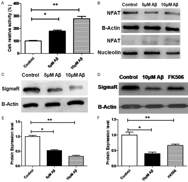
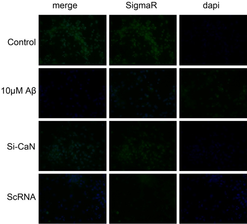
Sigma receptor is an endoplasmic reticulum protein and belongs to non-opioid receptor. Increasing evidence shows that Sigma receptor activation can significantly attenuate AD induced neurological dysfunction and the functional deficiency of Sigma receptor plays an important role in the Aβ induced neuronal loss. This study aimed to investigate the influence of extracellular accumulation of Aβ on the Sigma receptor expression. Our results showed the increase in extracellular Aβ had little influence on the mRNA expression of Sigma receptor, but gradually reduced its protein expression. Co-immunoprecipitation was employed to evaluate the interaction of Sigma receptor with other proteins. Results showed BIP could bind to Sigma receptor to affect the ubiquitination of Sigma receptor. Further investigation showed there was a NFAT binding site at the promoter of BIP. Then, Western blot assay was performed to detect NFAT expression. Results showed extracellular Aβ affected the nuclear translocation of NFAT and the CaN activity of NFAT also increased with the accumulation of extracellular Aβ. In this study, NFAT-BIP luciferase reporter gene system was constructed. Results showed NFAT was able to regulate the transcription of BIP. Thus, we speculate that extracellular Aβ accumulation may activate CaN/NFAT signaling pathway to induce chaperone BIP expression, which results in Sigma receptor ubiquitination and its degradation.

Keywords: Alzheimer disease; calcineurin; nuclear factor of activated T cells; sigma receptor; β-amyloid.


Figures

Figure 1
Figure 1
25-35 activates CaN/NFAT pathway and reduces Sigma-1R/BIP expression in N2A cells. A: CaN activity after treatment with Aβ at different concentrations. B: NFAT protein expression in the cytoplasm and nucleus after treatment with Aβ at different concentrations. C: SigmaR protein expression after treatment with Aβ at different concentrations. D: Influence of CaN inhibitor FK506 on SigmaR expression in N2A cells after treatment with 10 uM Aβ. C, E: Optical density in Western blot assay; D, F: Optical density in Western blot assay. All data are expressed as means ± SD (n=5/each group). *P<0.05, **P<0.01.
Figure 3
Figure 3
Transcriptional regulation of BIP by NFAT. (A) ChIP-PCR was used to detect the binding site of NFAT in BIP promoter. NFAT1 specific antibody was used to precipitate the corresponding genomic DNA segments and then real-time PCR was done to amplify the binding segments of NFAT1. input: DNA segments that were broken by ultrasound and not immunoprecipitated, but amplified with 3 pairs of primers. IgG: IgG was used to immunoprecipitate DNA segments that were broken by ultrasound, followed by amplification with 3 pairs of primers. primer2 and primer3: after immunoprecipitation with anti-NFAT1 antibody, the binding segments were amplified with 3 pairs of primers. Primer 2 and primer 3 were designed for the amplification of binding site at -1623 bp and -941 bp in (B), respectively. (B) Prediction of NFAT binding site in the BIP promoter. (C) Mutation binding sites of luciferase expression vector. In the synthesis of BIP promoter, deletion mutation was designed at 3 binding sites. (D) Effect of NFAT on the transcriptional activity of BIP promoters. BIP promoters with different length were cloned and contained different binding sites of NFAT. In 293T cells, the BIP promoter was cotransfected with NFAT1 gene, and then luciferase activity was detected. (E) Effect of NFAT on the transcriptional activity of BIP promoters with mutation at different sites. In the synthesis of BIP promoter, the deletion mutation was designed at the NFAT binding site. In 293T cells, the BIP promoter was cotransfected with NFAT1 gene, and then luciferase activity was detected. All data are expressed as means ± SD (n=5/each group). *P<0.05, **P<0.01.
Figure 2
Figure 2
BiP/Sigma-1R complex influences Sigma-1R ubiquitination and degradation in N2A cells. A: BiP/Sigma-1R complex co-immunoprecipitation after treatment with Aβ at different concentrations. input: no beads and antibody after Aβ treatment; anti-BIP: binding protein in the presence of BIP antibody. Immunoprecipitation was performed with anti-BIP antibody and then Western blotting to detect the expression of BIP, SigmaR and E3 ligase in the complex. B: Effect of E3 ligase on SigmaR protein expression: E3 ligase inhibitor MG132 and E3 ligase specific siRNA were used to treat N2A cells after treatment with 10 uM Aβ, and then Western blotting was performed to investigate the influence of E3 ligase on SigmaR expression. B, C: Optical density in Western blotting. All data atre expressed as means ± SD (n=5/each group). *P<0.05, **P<0.01.
Figure 4
Figure 4
Effects of CaN/NFAT pathway on Sigma and BIP expression in N2A cells after Aβ25-35 treatment. A: Detection of mRNA expression. N2A cells were treated with both 10 uM Aβ and CaN specific siRNA. Normal cells served as a control and scramble ScRNA as a negative control. B: Detection of CaN activity. Specific siRNA was used to down-regulate CaN mRNA expression and then CaN activity was detected with corresponding kit. C: Western blotting was done to detect protein expression. After silencing of CaN expression, Western blotting was done to detect the protein expression of CaN, NFAT, BIP and SigmaR. C, D: Optical density was detected after Western blotting. All data are expressed as means ± SD (n=5/each group). *P<0.05, **P<0.01.
Figure 5
Figure 5
Effects of CaN/NFAT pathway interfering on SigmaR expression in N2A cells. N2A cells were treated with both 10 uM Aβ and specific CaN siRNA. Normal cells served as a control and scramble ScRNA as a negative control. After 48-h treatment, fluorescence intensity was measured. Blue: Dapi positive nucleus; Green: SigmaR.

References

    1. Flemming A. Alzheimer’s disease: Abeta-fibrinogen interaction inhibitor improves cognition in AD. Nat Rev Drug Discov. 2014;13:494. - PubMed
    1. Geylis V, Steinitz M. Immunotherapy of Alzheimer’s disease (AD): from murine models to anti-amyloid beta (Abeta) human monoclonal antibodies. Autoimmun Rev. 2006;5:33–39. - PubMed
    1. Inoue S. In situ Abeta pores in AD brain are cylindrical assembly of Abeta protofilaments. Amyloid. 2008;15:223–233. - PubMed
    1. Robakis NK. Mechanisms of AD neurodegeneration may be independent of Abeta and its derivatives. Neurobiol Aging. 2011;32:372–379. - PMC - PubMed
    1. Villeneuve S, Reed BR, Madison CM, Wirth M, Marchant NL, Kriger S, Mack WJ, Sanossian N, DeCarli C, Chui HC, Weiner MW, Jagust WJ. Vascular risk and Abeta interact to reduce cortical thickness in AD vulnerable brain regions. Neurology. 2014;83:40–47. - PMC - PubMed

LinkOut - more resources