

Immunofluorescence on paraffin embedded renal biopsies: Experience of a tertiary care center with review of literature
- PMID: 27648410
- PMCID: PMC5011253
- DOI: 10.5527/wjn.v5.i5.461
Immunofluorescence on paraffin embedded renal biopsies: Experience of a tertiary care center with review of literature
Abstract
Aim: To describe the technique of immunofluorescence on paraffin embedded tissue sections and discuss the potential pitfalls with an in depth review of literature.
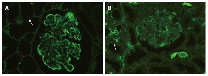
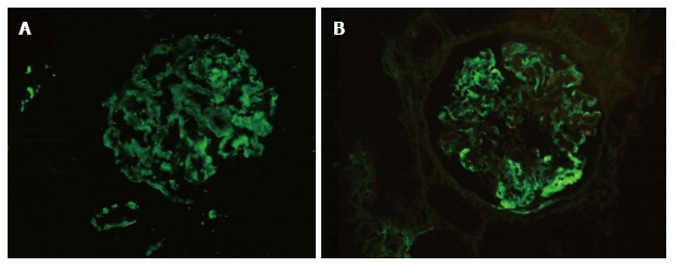
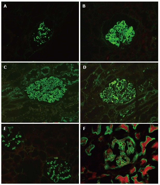
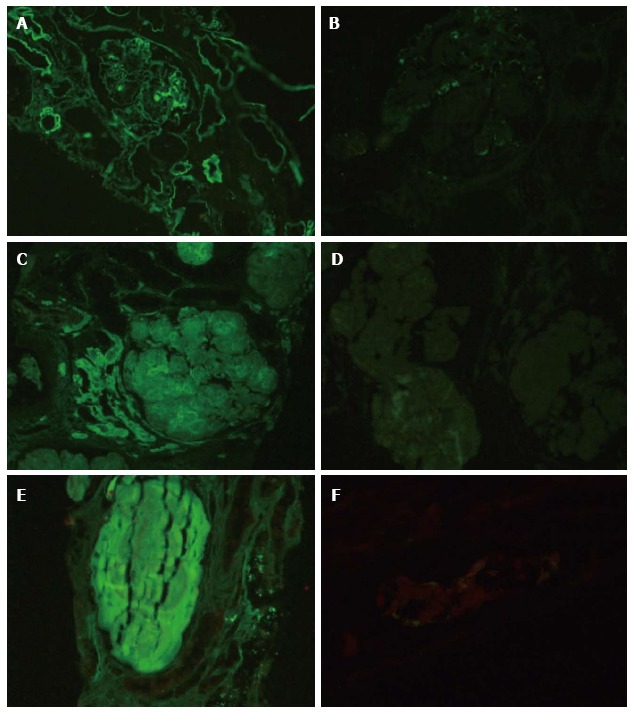
Methods: Immunofluorescence is integral to diagnostic renal pathology. Immunofluorescence on paraffin embedded renal biopsies (IF-P) after enzyme treatment has been described in literature, however has not found widespread use in renal pathology laboratories. In our laboratory proteinase K digestion of paraffin embedded renal biopsy material was standardized and applied prospectively in cases where immunofluorescence on fresh frozen tissue was non contributory or not possible. Diagnostic utility was assessed and in a cohort of cases comparison of intensity of staining with routine immunofluorescence was performed.
Results: Over the 5-year study period, of the 3141 renal biopsies received IF-P was performed on 246 cases (7.7%) and was interpretable with optimal digestion in 214 cases (6.8%). It was of diagnostic utility in the majority of cases, which predominantly included glomerular disease. Non-diagnostic IF-P was found in membranous nephropathy (2 of 11 cases), membranoproliferative glomerulonephritis (2 of 32 cases), lupus nephritis (1 of 25 cases), post infectious glomerulonephritis (1 of 11 cases) and chronic glomerulonephritis (3 of 8 cases). Comparing cases with both routine IF and IF-P, 35 of 37 showed either equal intensity or a minor difference in intensity of staining (1+) for the diagnostic immunoglobulin/complement. Technically assessment of immunofluorescence on the paraffin embedded tissue was found to be easier with clearly observed morphology, however a false positive staining pattern was observed in under-digested tissue.
Conclusion: As a "salvage" technique, immunofluorescence on paraffin embedded renal biopsies is of great diagnostic utility, however not without pitfalls.
Keywords: Enzymatic digestion; Immunofluorescence on paraffin section; Proteinase K; Renal biopsy; Salvage technique.
Figures
Similar articles
-
Paraffin Immunofluorescence: A Valuable Ancillary Technique in Renal Pathology.Kidney Int Rep. 2018 Jul 7;3(6):1260-1266. doi: 10.1016/j.ekir.2018.07.008. eCollection 2018 Nov. Kidney Int Rep. 2018. PMID: 30450452 Free PMC article. Review.
-
A Comparative Study of Immunofluorescence on Formalin-Fixed, Paraffin-Embedded Versus Fresh Frozen Kidney Biopsy.Cureus. 2023 Jun 26;15(6):e40978. doi: 10.7759/cureus.40978. eCollection 2023 Jun. Cureus. 2023. PMID: 37503479 Free PMC article.
-
Unmasking of complements using proteinase-K in formalin fixed paraffin embedded renal biopsies.Indian J Nephrol. 2016 May-Jun;26(3):182-7. doi: 10.4103/0971-4065.159558. Indian J Nephrol. 2016. PMID: 27194832 Free PMC article.
-
Diagnostic Utility of Immunofluorescence on Formalin-Fixed Paraffin-Embedded Skin Biopsy.Indian Dermatol Online J. 2024 Jun 10;15(4):620-623. doi: 10.4103/idoj.idoj_586_23. eCollection 2024 Jul-Aug. Indian Dermatol Online J. 2024. PMID: 39050059 Free PMC article.
-
IgG Subclass Staining in Routine Renal Biopsy Material.Am J Surg Pathol. 2016 May;40(5):617-26. doi: 10.1097/PAS.0000000000000605. Am J Surg Pathol. 2016. PMID: 26848798 Review.
Cited by
-
Monoclonal Gammopathy of Renal Significance: Histomorphological Spectrum at a Tertiary Care Center.Glomerular Dis. 2022 Aug 4;2(4):153-163. doi: 10.1159/000526244. eCollection 2022. Glomerular Dis. 2022. PMID: 36817291 Free PMC article.
-
Pitfalls and Caveats in Applying Chromogenic Immunostaining to Histopathological Diagnosis.Cells. 2021 Jun 15;10(6):1501. doi: 10.3390/cells10061501. Cells. 2021. PMID: 34203756 Free PMC article. Review.
-
Paraffin Immunofluorescence: A Valuable Ancillary Technique in Renal Pathology.Kidney Int Rep. 2018 Jul 7;3(6):1260-1266. doi: 10.1016/j.ekir.2018.07.008. eCollection 2018 Nov. Kidney Int Rep. 2018. PMID: 30450452 Free PMC article. Review.
-
A case of lupus vasculopathy presenting favorable renal outcome.CEN Case Rep. 2020 Feb;9(1):74-80. doi: 10.1007/s13730-019-00431-2. Epub 2019 Dec 20. CEN Case Rep. 2020. PMID: 31863345 Free PMC article.
-
Spectrum of Morphological and Immunofluorescence Patterns in Lupus Nephritis: A Single Institutional Study.Cureus. 2022 May 26;14(5):e25363. doi: 10.7759/cureus.25363. eCollection 2022 May. Cureus. 2022. PMID: 35765398 Free PMC article.
References
-
- Hed J, Eneström S. Detection of immune deposits in glomeruli: the masking effect on antigenicity of formalin in the presence of proteins. J Immunol Methods. 1981;41:57–62. - PubMed
-
- Huang SN, Minassian H, More JD. Application of immunofluorescent staining on paraffin sections improved by trypsin digestion. Lab Invest. 1976;35:383–390. - PubMed
-
- Nambirajan A, Bhowmik D, Singh G, Agarwal SK, Dinda AK. Monoclonal gammopathy of renal significance with light-chain deposition disease diagnosed postrenal transplant: a diagnostic and therapeutic challenge. Transpl Int. 2015;28:375–379. - PubMed
-
- Rheinhardt JM, Finkbeiner WE. Protease XXIV increases detection of mucin gene expression during in situ hybridization in archival tissue. J Histochem Cytochem. 2001;49:923–924. - PubMed
-
- Qualman SJ, Keren DF. Immunofluorescence of deparaffinized, trypsin-treated renal tissues. Preservation of antigens as an adjunct to diagnosis of disease. Lab Invest. 1979;41:483–489. - PubMed
LinkOut - more resources
Full Text Sources
Other Literature Sources
Research Materials
Miscellaneous
