Review
. 2016 Nov 23:50:175-210.
doi: 10.1146/annurev-genet-120215-035111. Epub 2016 Sep 14.

Control of Meiotic Crossovers: From Double-Strand Break Formation to Designation

Affiliations
Review

Control of Meiotic Crossovers: From Double-Strand Break Formation to Designation

Stephen Gray et al. Annu Rev Genet. .

Abstract

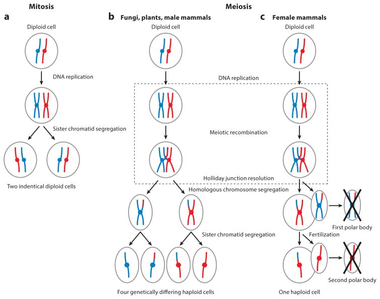
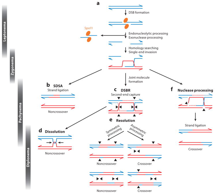
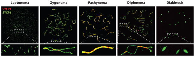
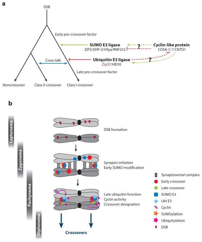
Meiosis, the mechanism of creating haploid gametes, is a complex cellular process observed across sexually reproducing organisms. Fundamental to meiosis is the process of homologous recombination, whereby DNA double-strand breaks are introduced into the genome and are subsequently repaired to generate either noncrossovers or crossovers. Although homologous recombination is essential for chromosome pairing during prophase I, the resulting crossovers are critical for maintaining homolog interactions and enabling accurate segregation at the first meiotic division. Thus, the placement, timing, and frequency of crossover formation must be exquisitely controlled. In this review, we discuss the proteins involved in crossover formation, the process of their formation and designation, and the rules governing crossovers, all within the context of the important landmarks of prophase I. We draw together crossover designation data across organisms, analyze their evolutionary divergence, and propose a universal model for crossover regulation.

Keywords: crossover designation; homologous recombination; meiosis.


Figures

Figure 1
Figure 1
Mitosis and meiosis. (a) During mitosis, a diploid cell containing a pair of homologous chromosomes (one red and one blue) undergoes DNA replication to generate sister chromatids. In the mitotic division, sister chromatids are segregated to separate daughter cells. The final products of mitosis are two genetically identical cells. As in mitosis, (b) in meiosis, diploid cells containing two homologous chromosomes undergo DNA replication to generate sister chromatids. During prophase I (dotted box), however, meiotic cells then undergo recombination, forming physical links between homologs and resulting in genetic exchange. In the first meiotic division, homologous chromosomes are segregated, followed by sister chromatids in the second division. The products of meiosis in fungi, plants, and male mammals are four genetically differing haploid gametes from one starting diploid cell. (c) In female meiosis in mammals, cells arrest prior to the first meiotic division (dictyate arrest). Upon ovulation, the first meiotic division occurs, segregating homologous chromosomes and forming a polar body, which is subsequently discarded. Upon fertilization, segregation of the sister chromatids occurs during the second meiotic division, creating a second polar body, which is also subsequently discarded. The product of female meiosis is one genetically differing haploid cell.
Figure 2
Figure 2
Summary of meiotic recombination based on data from Saccharomyces cerevisiae. (a) Meiotic recombination initiated by the formation of DNA double-strand breaks (DSBs) by the protein Spo11 (orange ellipse) and other accessory proteins. Following DNA cleavage downstream of the DSB event, Spo11 is liberated, forming a Spo11-oligonucleotide complex. Exonuclease activity generates single-stranded DNA (ssDNA) that, through the action of the RecA homologs Rad51 and Dmc1, coat the ssDNA, invade homologous DNA templates on the homolog, and create a displacement (D)-loop. (b) In the following extension of the invading DNA strand, the strand can become disrupted and displaced, reanneal with the opposite side of the DSB, and be repaired as a noncrossover event in a process known as synthesis-dependent strand annealing (SDSA). Alternatively, the invading strand can continue to become extended creating a larger D-loop. (c) Upon second-end capture of the other side of the DSB and subsequent DNA synthesis, a double Holliday junction (dHJ) is formed. (d) By the actions of a helicase and a topoisomerase, dHJs can be migrated toward each other and dissolved (in a process called dissolution) into a noncrossover event. (e) Alternatively, dHJs can be resolved in a symmetrical manner creating noncrossover events or asymmetrically creating crossover events. (f) Following strand invasion, structure-specific nucleases can process joint molecule intermediates to generate a crossover. Many of these events are drawn largely from studies in S. cerevisiae and can be extrapolated to many meiotic species. Abbreviation: DSRB, double-strand break repair.
Figure 3
Figure 3
Synaptonemal complex (SC) formation and synapsis during prophase I in mouse. Meiotic chromosome spread preparations from male mouse spermatocytes are immunofluorescently stained and imaged at different stages of prophase I using 3D structured illumination microscopy (3D-SIM). During prophase I, axial elements [stained with antibodies against SYCP3 (green)] start to form along the homologous chromosomes during leptonema. In zygonema, axial elements have formed along the entire axis of the homologous chromosome, and as synapsis occurs proteins of the central/transverse filament [stained with antibodies against SYCP1 (red)] begin to zipper the homologous chromosomes together. In pachynema, full synapsis has occurred along all autosomes and within the pseudoautosomal region of the sex chromosomes (in males). During diplonema, the SC starts to disassemble and homologous chromosomes repel each other, except for at regions of crossovers. Finally, at diakinesis, the axial elements are visible only at the centromeres.
Figure 4
Figure 4
Model for crossover control. (a) The interaction between double-strand break (DSB) repair pathways is orchestrated by SUMO and ubiquitin E3 ligases and overseen by cyclin-like proteins such as COSA-1/CNTD1. DSBs can repair as either noncrossovers or crossovers, with cross talk occurring between the crossover classes (blue arrow). Three groups of proteins have been described as affecting crossover control: SUMO E3 ligases, ubiquitin E3 ligases, and cyclin-like proteins. All three proteins function in the process of paring down early crossover promoting factors to late crossover promoting factors. SUMO E3 ligases likely function to select/determine/designate some pro-crossover sites. Given that the mitotic role of HEI10 is inhibition of related cyclin-like proteins, it is likely that the ubiquitin E3 ligases inhibit cyclin-like proteins in meiosis and target some pro-crossover sites. It is unknown how the cyclin-like proteins function in relation to the SUMO E3 ligases. (b) Spatiotemporal relationship between SUMO and ubiquitin E3 ligases. The role of synapsis initiation sites and RING-finger proteins are depicted.
Figure 5
Figure 5
Divergence of crossover control mechanisms. Phylogenetic tree of species divergence indicates known meiotic crossover designation proteins (green) with mitotic equivalents (if known; blue). Proteins in purple are predicted or currently unknown proteins. Predicted crossover control mechanisms and their divergences are marked in red.

References

    1. Agarwal S, Roeder GS. Zip3 provides a link between recombination enzymes and synaptonemal complex proteins. Cell. 2000;102(2):245–55. - PubMed
    1. Allers T, Lichten M. Intermediates of yeast meiotic recombination contain heteroduplex DNA. Mol Cell. 2001;8(1):225–31. - PubMed
    1. Allers T, Lichten M. Differential timing and control of noncrossover and crossover recombination during meiosis. Cell. 2001;106(1):47–57. - PubMed
    1. Anderson CM, Oke A, Yam P, Zhuge T, Fung JC. Reduced crossover interference and increased zmm-independent recombination in the absence of Tel1/ATM. PLOS Genet. 2015;11(8):e1005478. - PMC - PubMed
    1. Anderson EL, Baltus AE, Roepers-Gajadien HL, Hassold TJ, de Rooij DG, et al. Stra8 and its inducer, retinoic acid, regulate meiotic initiation in both spermatogenesis and oogenesis in mice. PNAS. 2008;105(39):14976–80. - PMC - PubMed

LinkOut - more resources