. 2016 Sep 20;11(9):e0163100.
doi: 10.1371/journal.pone.0163100. eCollection 2016.

HIV-1 Vpr Abrogates the Effect of TSG101 Overexpression to Support Virus Release

Affiliations

HIV-1 Vpr Abrogates the Effect of TSG101 Overexpression to Support Virus Release

Nopporn Chutiwitoonchai et al. PLoS One. .

Abstract

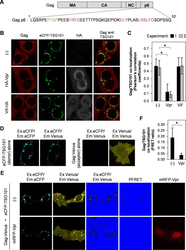
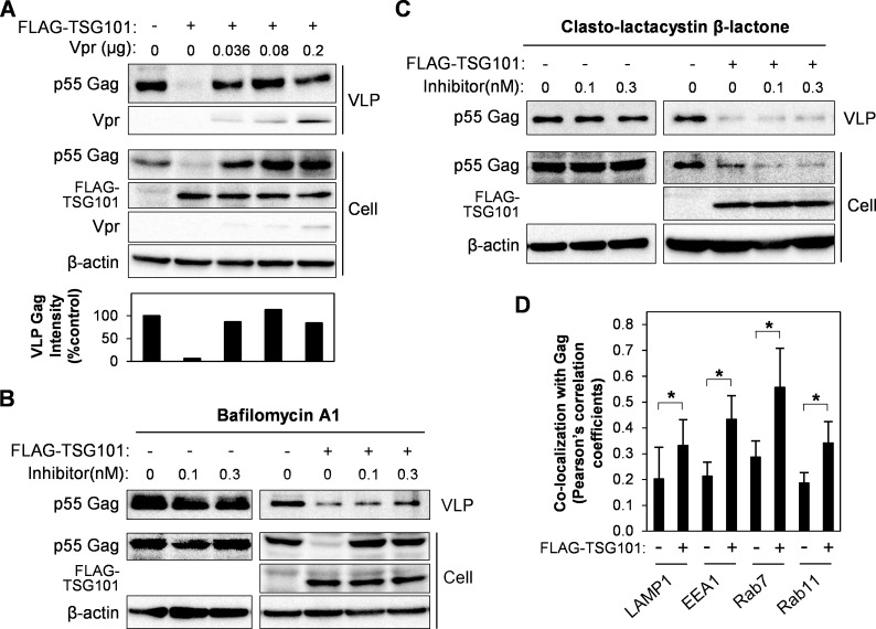
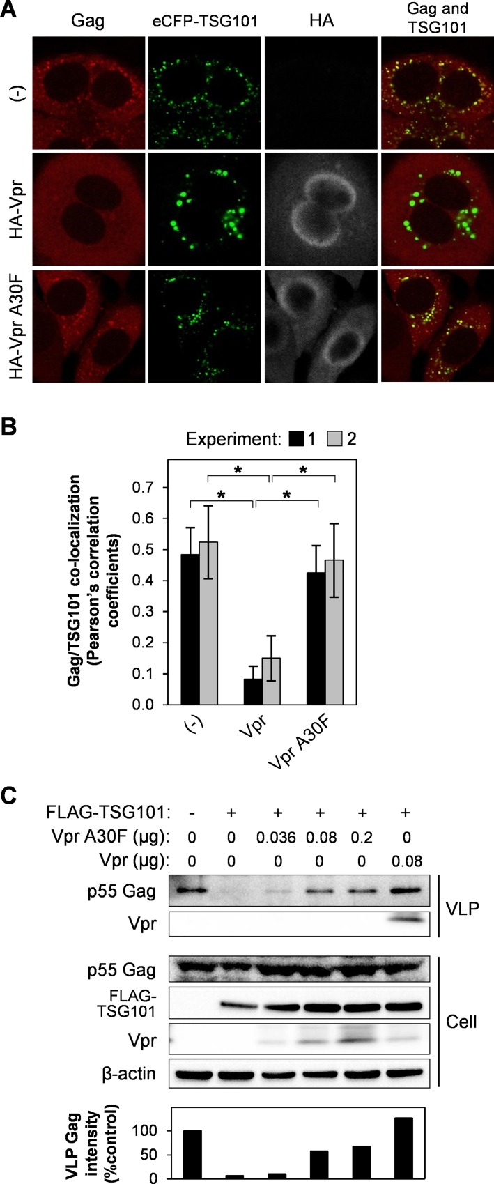
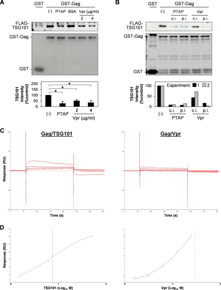
HIV-1 budding requires interaction between Gag and cellular TSG101 to initiate viral particle assembly and release via the endosomal sorting complexes required for transport (ESCRT) pathway. However, some reports show that overexpression of TSG101 inhibits virus release by disruption of Gag targeting process. Since a HIV-1 accessory protein, Vpr binds to Gag p6 domain at the position close to the binding site for TSG101, whether Vpr implicates TSG101 overexpression effect has not been investigated. Here, we found that Vpr abrogates TSG101 overexpression effect to rescue viral production. Co-transfection of TSG101 and Gag with Vpr prevented TSG101-induced Gag accumulation in endosomes and lysosomes. In addition, Vpr rescued virus-like particle (VLP) production in a similar manner as a lysosomal inhibitor, Bafilomycin A1 indicating that Vpr inhibits TSG101-induced Gag downregulation via lysosomal pathway. Vpr and Gag interaction is required to counteract TSG101 overexpression effect since Vpr A30F mutant which is unable to interact with Gag and incorporate into virions, reduced ability to prevent Gag accumulation and to rescue VLP production. In addition, GST pull-down assays and Biacore analysis revealed that Vpr competed with TSG101 for Gag binding. These results indicate that Vpr overcomes the effects of TSG101 overexpression to support viral production by competing with TSG101 to bind Gag.


Conflict of interest statement

The authors have declared that no competing interests exist.

Figures

Fig 1
Fig 1. Vpr prevents TSG101-induced Gag accumulation at perinuclear region.
(A) Schematic showing the binding motifs for TSG101 (PTAP; green) and Vpr (FRFG, ELY, and LXSLFG; red) within the Gag p6 domain. (B) HeLa cells were co-transfected with 0.8 μg of pCAGGS/Gag and 1.5 μg of pCAGGS/eCFP-TSG101 either without/with 0.5 μg of pCAGGS/HA-Vpr or 0.5 μg of pCAGGS/Vif-HA for 48 h prior to immunofluorescence staining with an anti-Gag and anti-HA antibodies, followed by an Alexa Fluor 594 goat anti-rabbit antibody and an Alexa Fluor 633 goat anti-mouse antibody. Image acquisition was performed under a confocal laser-scanning microscope. (C) Co-localization of Gag/TSG101 was analyzed by Pearson’s correlation coefficients. Data represent the means ± SD of the result of two independent experiments. *, P < 0.05 (unpaired t-test). (D-E) HeLa cells were co-transfected with 1.5 μg of pCAGGS/eCFP-TSG101 or 0.8 μg of pCAGGS/Gag-Venus (D) or 1.5 μg of pCAGGS/eCFP-TSG101 and 0.8 μg of pCAGGS/Gag-Venus either without/with 0.5 μg of pCAGGS/mRFP-Vpr (E) for 48 h before fixation and image acquisition. The precision FRET (PFRET) signal was analyzed using the sensitized emission method. The FRET (Ex.eCFP/Em.Venus) and PFRET image colors were converted by Hi/Lo function of FV10-ASW v.2.1 software (Olympus) to facilitate visualization of Gag/TSG101 co-localization. (F) FRET ratio (PFRET signal divided by the donor eCFP-TSG101 signal) of Gag/TSG101 co-localization in the absence or presence of Vpr. Data represent means ± SD of the result of one representative experiment from two independent performs. *, P < 0.05 (unpaired t-test).
Fig 2
Fig 2. Vpr rescues VLP production by inhibition of TSG101-induced Gag degradation via lysosomal pathway.
(A) HEK293T cells were transfected with 0.8 μg of pCAGGS/Gag, or with 0.8 μg of pCAGGS/Gag plus 1.5 μg of pCAGGS/FLAG-TSG101 either without/with different amounts of pCAGGS/Vpr for 48 h. VLPs in the cultured medium were collected by a 20% sucrose cushion. Whole cell lysates were prepared and samples were subjected to western blot analysis with anti-Gag, anti-Vpr, anti-FLAG, and anti-β-actin antibodies. The bottom panel represents intensity of VLP Gag from western blot analysis. Data represent the result of one representative experiment from three independent performs. (B-C) HEK293T cells were transfected with 0.8 μg of pCAGGS/Gag, or with 0.8 μg of pCAGGS/Gag and 1.5 μg of pCAGGS/FLAG-TSG101, for 24 h, before addition of Bafilomycin A1 (B) or Clasto-lactacystin β-lactone (C), followed by further culture for 16 h. VLPs and whole cell lysates were prepared and subjected to western blot analysis with anti-Gag, anti-Vpr, anti-FLAG, and anti-β-actin antibodies. Data represent the result of one representative experiment from two independent performs. (D) HeLa cells were transfected with 0.8 μg of pCAGGS/Gag-Venus, or with 0.8 μg of pCAGGS/Gag-Venus plus 1.5 μg of pCAGGS/eCFP-TSG101, for 48 h, followed by immunofluorescence staining anti-LAMP1, anti-EEA1, anti-Rab7, or anti-Rab11 antibody with a secondary antibody, Alexa Fluor 594 goat anti-rabbit antibody. Co-localization of Gag/LAMP1, Gag/EEA1, Gag/Rab7, or Gag/Rab11 was analyzed by Pearson’s correlation coefficients. Data represent means ± SD of the result of one representative experiment. *, P < 0.05 (unpaired t-test).
Fig 3
Fig 3. Vpr and Gag interaction is required to abrogate the effect of TSG101 overexpression.
(A) HeLa cells were co-transfected with 0.8 μg of pCAGGS/Gag and 1.5 μg of pCAGGS/eCFP-TSG101 either without/with 0.5 μg of pCAGGS/HA-Vpr or pCAGGS/HA-Vpr A30F for 48 h prior to immunofluorescence staining with an anti-Gag and anti-HA antibodies, followed by an Alexa Fluor 594 goat anti-rabbit antibody and an Alexa Fluor 633 goat anti-mouse antibody. Co-localization of Gag/TSG101 was analyzed by Pearson’s correlation coefficients (B). Data represent the means ± SD of the result of two independent experiments. *, P < 0.05 (unpaired t-test). (C) HEK293T cells were transfected with 0.8 μg of pCAGGS/Gag, or with 0.8 μg of pCAGGS/Gag plus 1.5 μg of pCAGGS/FLAG-TSG101 either without/with different amounts of pCAGGS/Vpr A30F or 0.08 μg of pCAGGS/Vpr (positive control). After 48 h, VLPs in the cultured medium were collected by a 20% sucrose cushion. Whole cell lysates were prepared and samples were subjected to western blot analysis with anti-Gag, anti-Vpr, anti-FLAG, and anti-β-actin antibodies. The bottom panel represents intensity of VLP Gag from western blot analysis. Data represent the result of one representative experiment from two independent performs.
Fig 4
Fig 4. Vpr rescues the TSG101-mediated inhibition of viral particle release.
(A) HeLa cells were transfected with 0.3 μg of pNL43 Luc E- R+ (Vpr+) or pNL43 Luc E- R- (Vpr-) either without/with 0.1 μg of pCAGGS/mRFP-TSG101 and 0.1 μg of pcDNA/YFP-Vpr for 16 h prior to immunofluorescence staining with an anti-Gag AG3.0 antibody followed by a FITC-conjugated sheep anti-mouse antibody. White arrows indicate Gag/TSG101 co-localized on/near the plasma membrane observed by total internal reflection fluorescence (TIRF) microscopy. Data represents the result of one representative experiment from two independent performs. (B-C) HEK293T cells were transfected with 0.2 μg of pNL43 Luc E- R+ (Vpr+) (B) or pNL43 Luc E- R- (Vpr-) (C), together with 1 μg of pCAGGS/FLAG-TSG101 and different amounts of pCAGGS/Vpr for 48 h. Viral particles in the culture medium were collected by PEG precipitation. Whole cell lysates were prepared and samples were subjected to western blotting with anti-Gag, anti-Vpr, anti-FLAG, and anti-β-actin antibodies. Data represents the result of one representative experiment from two independent performs.
Fig 5
Fig 5. Vpr competes with TSG101 for binding to Gag.
(A) GST or GST-Gag protein were immobilized on Glutathione Sepharose beads prior to overnight incubation with purified FLAG-TSG101 protein in the absence/presence of 48.3 μg/ml of PTAP short peptide (a positive control which targets the UEV domain of TSG101), 48.3 μg/ml of BSA (negative control), or 2.0 or 4.0 μg/ml of purified HA-Vpr protein. The beads were pulled-down, washed, and subjected to western blotting with anti-FLAG and anti-GST antibodies. The bottom panel represents mean ± SD intensity values of Gag-bound TSG101 from western blot analysis of four independent performs. *, P < 0.05 (unpaired t-test). (B) GST-Gag was co-incubated with purified FLAG-TSG101 and 48.3 μg/ml of PTAP short peptide or 2 μg/ml of HA-Vpr protein (competitive incubation, c.i.), or allowed to bind with PTAP short peptide or HA-Vpr protein for 3 h before co-incubation with FLAG-TSG101 protein (pre-incubation, p.i.). After overnight incubation, the beads were pulled-down, washed, and subjected to western blotting with anti-FLAG and anti-GST antibodies. The bottom panel represents intensity of Gag-bound TSG101 from western blot analysis of two independent performs. (C-D) GST-Gag protein was immobilized on Biacore sensor chip CM5 and the surface was injected with purified FLAG-TSG101 or FLAG-Vpr protein at different concentrations by Biacore T100 instrument. Kinetic sensorgrams of Gag-bound TSG101 at 0, 15.6, 31.3, 62.5, and 500.0 nM (C, left panel) and Gag-bound Vpr at 0, 15.6, 31.3, 62.5, 125.0, and 250.0 nM (C, right panel) are showed. (D) Steady stage affinity fitting curves of Gag/TSG101 (left panel) and Gag/Vpr (right panel) binding at equilibrium (4 s before injection stop). Black and red vertical lines represent equilibrium dissociation constant (KD) of Gag/TSG101 and Gag/Vpr binding affinity, representatively. Data represents the result of one representative experiment from two independent performs.

Similar articles

Cited by

References

    1. Bell NM, Lever AM. HIV Gag polyprotein: processing and early viral particle assembly. Trends Microbiol. 2013;21(3):136–44. Epub 2012/12/26. 10.1016/j.tim.2012.11.006 . - DOI - PubMed
    1. Freed EO. HIV-1 gag proteins: diverse functions in the virus life cycle. Virology. 1998;251(1):1–15. Epub 1998/11/14. 10.1006/viro.1998.9398 . - DOI - PubMed
    1. Garrus JE, von Schwedler UK, Pornillos OW, Morham SG, Zavitz KH, Wang HE, et al. Tsg101 and the vacuolar protein sorting pathway are essential for HIV-1 budding. Cell. 2001;107(1):55–65. Epub 2001/10/12. . - PubMed
    1. Martin-Serrano J, Zang T, Bieniasz PD. HIV-1 and Ebola virus encode small peptide motifs that recruit Tsg101 to sites of particle assembly to facilitate egress. Nat Med. 2001;7(12):1313–9. Epub 2001/12/01. 10.1038/nm1201-1313 . - DOI - PubMed
    1. VerPlank L, Bouamr F, LaGrassa TJ, Agresta B, Kikonyogo A, Leis J, et al. Tsg101, a homologue of ubiquitin-conjugating (E2) enzymes, binds the L domain in HIV type 1 Pr55(Gag). Proc Natl Acad Sci U S A. 2001;98(14):7724–9. Epub 2001/06/28. 10.1073/pnas.131059198 - DOI - PMC - PubMed

MeSH terms

Substances