. 2016 Sep 21:6:33493.
doi: 10.1038/srep33493.

Quantitative Analysis of the Microtubule Interaction of Rabies Virus P3 Protein: Roles in Immune Evasion and Pathogenesis

Affiliations

Quantitative Analysis of the Microtubule Interaction of Rabies Virus P3 Protein: Roles in Immune Evasion and Pathogenesis

Aaron Brice et al. Sci Rep. .

Abstract

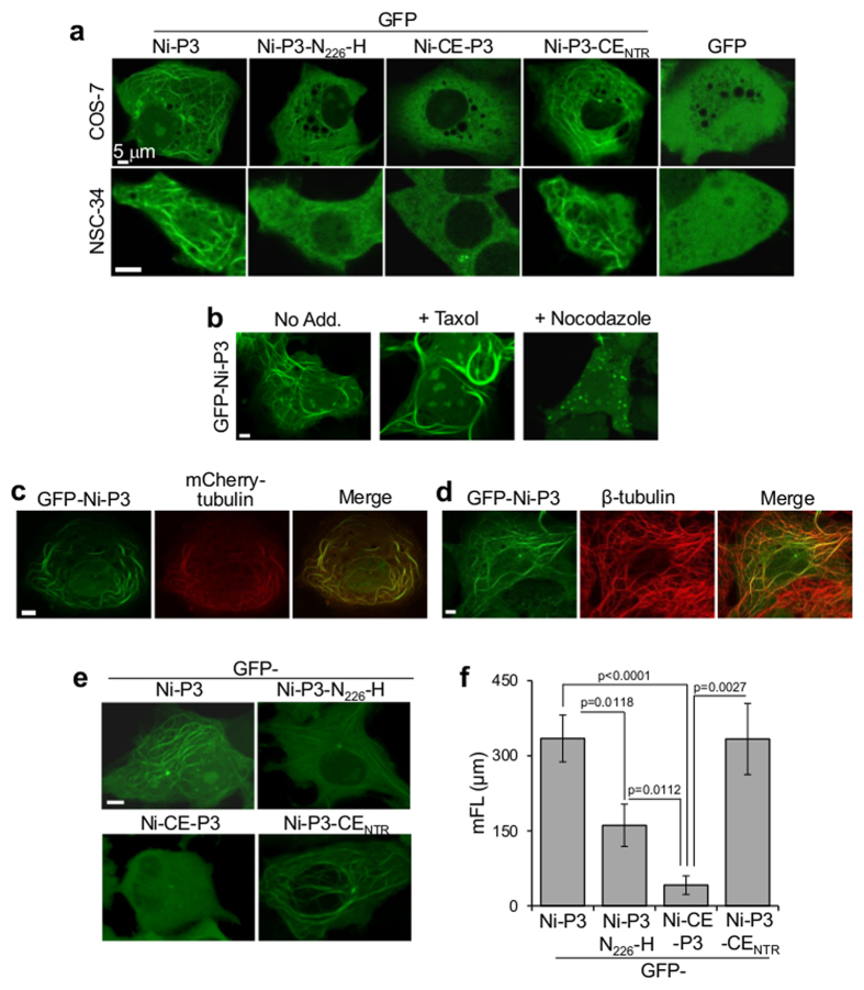
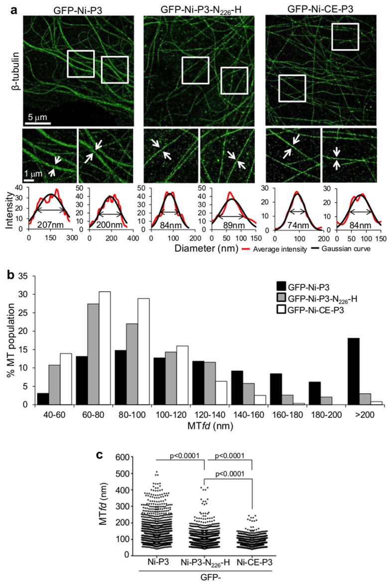
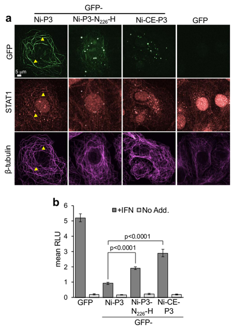
Although microtubules (MTs) are known to have important roles in intracellular transport of many viruses, a number of reports suggest that specific viral MT-associated proteins (MAPs) target MTs to subvert distinct MT-dependent cellular processes. The precise functional importance of these interactions and their roles in pathogenesis, however, remain largely unresolved. To assess the association with disease of the rabies virus (RABV) MAP, P3, we quantitatively compared the phenotypes of P3 from a pathogenic RABV strain, Nishigahara (Ni) and a non-pathogenic Ni-derivative strain, Ni-CE. Using confocal/live-cell imaging and dSTORM super-resolution microscopy to quantify protein interactions with the MT network and with individual MT filaments, we found that the interaction by Ni-CE-P3 is significantly impaired compared with Ni-P3. This correlated with an impaired capacity to effect association of the transcription factor STAT1 with MTs and to antagonize interferon (IFN)/STAT1-dependent antiviral signaling. Importantly, we identified a single mutation in Ni-CE-P3 that is sufficient to inhibit MT-association and IFN-antagonist function of Ni-P3, and showed that this mutation alone attenuates the pathogenicity of RABV. These data provide evidence that the viral protein-MT interface has important roles in pathogenesis, suggesting that this interface could provide targets for vaccine/antiviral drug development.


Figures

Figure 1
Figure 1. Schematic representation of P protein and the P3 proteins used in this study.
P3 is an N-terminally truncated isoform of P protein comprising residues 53–297, which is generated in infected cells by ribosomal leaky scanning that results in translation from an internal AUG codon corresponding to M53 (arrow) of the full length P protein (called P1). The CTD (containing the MT, N-RNA and STAT1 binding regions) and NTR (containing the dimerization domain) are indicated; residue positions are indicated beneath the P1 protein. Residues at positions 56, 58, 66, 81 and 226 differ between P3 from the pathogenic (Ni) and attenuated (Ni-CE) strains of RABV (substitutions in Ni-CE-P3 are in red).
Figure 2
Figure 2. Interaction of Ni-P3 with MTs is impaired by N226-H mutation.
(a) COS-7 cells were transfected to express the indicated proteins before analysis by live-cell CLSM; each image is representative of cells in 30 fields of view sampled over 3 separate assays (COS-7) or 9 fields of view (NSC-34). (b–d) COS-7 cells transfected to express GFP-Ni-P3 were treated with or without Taxol or nocodazole (b) co-transfected to express mCherry-tubulin (c) or fixed and immunostained for β-tubulin (d) before analysis by CLSM; colocalization in b and d is apparent as yellow coloration in merged image. (e) Live COS-7 cells expressing the indicated proteins were analyzed by CLSM to generate deconvoluted 3D images (images show reconstructed 3D images viewed down the z-axis). (f) Images such as those shown in (e) were analyzed to derive mean filament length values (mFL ± SEM; n ≥ 30 cells from 3 identical assays). p values were determined using the Mann Whitney test.
Figure 3
Figure 3
N226-H mutation inhibits colocalization of P3 with MTs (a) COS-7 cells expressing the indicated proteins were extracted to remove soluble (non-MT-associated) protein before fixation and immunostaining for β-tubulin, and analysis by CLSM (colocalization is apparent as white coloration in merged image). (b) Images such as those shown in (a) were analyzed to derive Pearson’s coefficient as a measure of colocalization between GFP-P3 and β-tubulin (mean ± SEM; n ≥ 14 cells from two assays). p values were determined using Student’s t-test or the Mann-Whitney test.
Figure 4
Figure 4. Quantitative dSTORM analysis of P3-induced bundling of MTs.
(a) dSTORM images of immunostained β-tubulin in COS-7 cells expressing the indicated proteins are shown in the upper panels. Boxed areas are expanded in the lower panels, and Gaussian functions (black line) fit to the average intensity profile (red line) of the indicated filaments shown below. The derived MTfd (calculated at the full width half-height of the Gaussian function) is indicated. (b) The frequency distribution of MTfds calculated for each protein is shown (n = 1294 [GFP-Ni-P3], 1071 [GFP-Ni-CE-P3] and 1299 [GFP-Ni-P3-N226-H]; measurements are from 10 cells for each protein over two identical assays). (c) Scatter dot plots of MTfds shown in (b). p values were determined using the Mann-Whitney test.
Figure 5
Figure 5. Ni-CE P can interact with N-protein and contains a functional homodimerisation region.
L40 yeast cells were cotransformed to express the DNA-binding domain of LexA (Lex) or the GAL4-activation domain (GAD) fused to Ni-P1, Ni-CE-P1, Ni-P3, Ni-CE P3, the corresponding P3 NTRs (P54-172), or RABV N protein. Growth streaks are shown, where interaction of Lex- and GAD-fused proteins is indicated by dark coloration of colonies.
Figure 6
Figure 6. N226-H mutation impairs the IFN-antagonistic function of P3.
(a) IFNα-treated COS-7 cells expressing the indicated proteins were extracted before fixation and immunostaining for STAT1 and β-tubulin, and analysis by CLSM; images are representative of 15 fields of view from two assays. (b) IFN-α-dependent signaling in COS-7 cells expressing the indicated proteins was analyzed using a dual luciferase reporter gene assay, as previously described. Luciferase activity is expressed as fold change relative to that obtained for IFN-α-treated cells expressing Ni-P3 protein (mean relative light units [RLU] ± SEM; n = 12 from 4 identical assays). p values were calculated using Student’s t-test.
Figure 7
Figure 7. N226-H mutation attenuates RABV.
(a) Schematic representation of the genomes of viruses used; genes from Ni and Ni-CE are in black and white, respectively. The P gene of Ni-CE is substituted for the P gene of Ni in CE(NiP), and for a mutated version of the Ni P gene containing N226-H in CE(NiP-N226-H). (b,c) 100 (b) or 106 (c) FFU of the indicated virus was inoculated intracerebrally into mice (five mice per condition) and body weight relative to that at 0 dpi (mean relative body weight ± SEM, for live mice) and survival was monitored over 21 or 14 dpi. p-values for survival curves were calculated using log-rank (Mantel-Cox) test. all mice dead or sacrificed on reaching end point.

Similar articles

Cited by

References

    1. Allan V. J. Cytoplasmic dynein. Biochemical Society transactions 39, 1169–1178, 10.1042/BST0391169 (2011). - DOI - PubMed
    1. de Forges H., Bouissou A. & Perez F. Interplay between microtubule dynamics and intracellular organization. The international journal of biochemistry & cell biology 44, 266–274, 10.1016/j.biocel.2011.11.009 (2012). - DOI - PubMed
    1. Hirokawa N., Niwa S. & Tanaka Y. Molecular motors in neurons: transport mechanisms and roles in brain function, development, and disease. Neuron 68, 610–638, 10.1016/j.neuron.2010.09.039 (2010). - DOI - PubMed
    1. Mimori-Kiyosue Y. Shaping microtubules into diverse patterns: molecular connections for setting up both ends. Cytoskeleton 68, 603–618, 10.1002/cm.20540 (2011). - DOI - PubMed
    1. Greber U. F. & Way M. A superhighway to virus infection. Cell 124, 741–754, 10.1016/j.cell.2006.02.018 (2006). - DOI - PubMed

Publication types