Review
. 2016 Nov;17(11):703-721.
doi: 10.1038/nrm.2016.111. Epub 2016 Sep 21.

Roles of eukaryotic topoisomerases in transcription, replication and genomic stability

Affiliations
Review

Roles of eukaryotic topoisomerases in transcription, replication and genomic stability

Yves Pommier et al. Nat Rev Mol Cell Biol. 2016 Nov.

Abstract

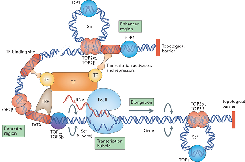
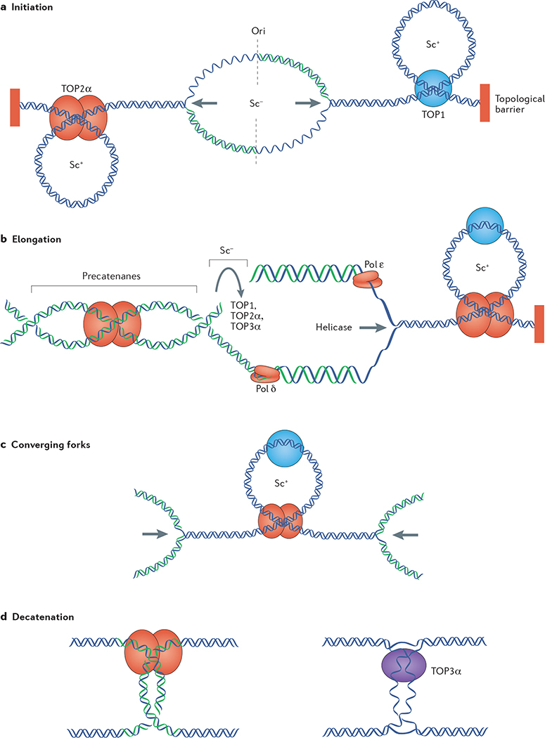
Topoisomerases introduce transient DNA breaks to relax supercoiled DNA, remove catenanes and enable chromosome segregation. Human cells encode six topoisomerases (TOP1, TOP1mt, TOP2α, TOP2β, TOP3α and TOP3β), which act on a broad range of DNA and RNA substrates at the nuclear and mitochondrial genomes. Their catalytic intermediates, the topoisomerase cleavage complexes (TOPcc), are therapeutic targets of various anticancer drugs. TOPcc can also form on damaged DNA during replication and transcription, and engage specific repair pathways, such as those mediated by tyrosyl-DNA phosphodiesterase 1 (TDP1) and TDP2 and by endonucleases (MRE11, XPF-ERCC1 and MUS81). Here, we review the roles of topoisomerases in mediating chromatin dynamics, transcription, replication, DNA damage repair and genomic stability, and discuss how deregulation of topoisomerases can cause neurodegenerative diseases, immune disorders and cancer.


Conflict of interest statement

Competing interests statement

The authors declare no competing interests.

Figures

Figure 1 |
Figure 1 |. Overview of eukaryotic topoisomerases.
a–c | Topoisomerases act by cleaving the DNA phosphodiester backbone and forming transient covalent linkages between a Tyr residue and the DNA 3′ end (TOP1 enzymes) or 5′ end (TOP2 and TOP3 enzymes). Resealing of the breaks is carried out by nucleophilic attack (arrows) of the 5′-hydroxyl end in the case of TOP1 enzymes and the 3′-hydroxyl end in the case of TOP2 and TOP3 enzymes. Base stacking (dashed double-headed arrows) is crucial for the realignment of the DNA ends (like a molecular zipper) and their re-ligation. d | TOP1 enzymes relax both negative and positive supercoils (Sc−/+) by nicking one strand and allowing controlled rotation of the broken strand around the intact strand. TOP1 enzymes can re-ligate non-homologous ends, thereby acting as DNA recombinases. e | TOP2 enzymes function as homodimers to relax both negative and positive supercoils and to resolve catenanes and DNA knots, explaining their essential role in cell division, during which supercoiled circles form catenated daughter molecules. They cleave both DNA strands with a four-base stagger, thereby directing a second duplex to pass through (duplex passage), and re-ligating the DNA following the passage. They require both Mg2+ and ATP hydrolysis for their catalytic cycle. f | TOP3 enzymes only relax hypernegative supercoiling (HSc) by cleaving one of the two strands of DNA in regions where negative supercoiling promotes their separation and by passing the intact strand through the broken one. Mg2+ is a required metal cofactor. TOP3β can act as an RNA helicase and resolve R loops. See Supplementary information S1 (box), for further information on DNA supercoiling.
Figure 2 |
Figure 2 |. Topoisomerases and transcription.
Transcription incurs topological constraints that result from the progression of RNA polymerase II (Pol II). Positive supercoiling (Sc+) of the DNA template takes place ahead of the transcription bubble, which in turn obstructs further Pol II movement, and negative supercoiling (Sc), which promotes the formation of RNA–DNA hybrids (R loops), accumulates behind it. TOP2 and especially TOP1 enzymes function ahead of Pol II to remove positive supercoils, whereas relaxation of negative supercoils behind the transcription apparatus relies on TOP1 and TOP3β. In addition, TOP1 regulates the activity of the transcription factor TATA-box-binding protein (TBP) at promoter TATA boxes independently of its catalytic activity. The formation of TOP2β-mediated transient DNA double-stranded breaks at promoter regions in certain genes is crucial for transcription activation. TOP1 is also recruited to certain enhancer regions to promote (ligand-dependent) enhancer activation by generating transient DNA single-stranded breaks. Topological barriers are genomic regions where the DNA is not free to rotate around its axis and require TOP1 and TOP2 to relax supercoils (Sc). TF, transcription factor.
Figure 3 |
Figure 3 |. Functions of topoisomerases in DNA replication.
a | Initiation of DNA replication requires separation of the two parental strands, which generates negative supercoiling (Sc) at the origin of replication «and positive supercoiling (Sc+) in the flanking regions owing to topological barriers, such as nuclear matrix attachment sites or insulators, where the DNA is not free to rotate around its axis. Positive supercoiling is dissipated by TOP1 and TOP2α to allow replication fork progression (indicated by the arrows). b | Replication elongation generates positive supercoiling ahead of the replication fork and negative supercoiling behind it. Positive supercoiling is removed by TOP1 and TOP2α, whereas negative supercoiling can be removed by TOP1, TOP2α or TOP3α. TOP2α can also remove precatenanes, which are formed when the fork rotates during elongation. c | Converging forks generate high positive supercoiling between them. d | Upon replication completion, catenanes are removed by TOP2α (left) and hemicatenanes are removed by TOP3α (right). Pol, DNA polymerase.
Figure 4 |
Figure 4 |. Topoisomerases and DNA damage.
a–c | Collision of a replication fork into a TOP1 cleavage complex (TOP1cc; part a) produces replication run-off (part b) in which newly replicated DNA (red) is extended by DNA polymerases up to the 5′ end of the broken DNA, thereby generating a double-stranded end (DSE), which is repaired by homologous recombination. Alternatively, replication fork reversal regenerates a reversible TOP1cc and produces a ‘chicken foot’ structure (part c). d–i | Processing of TOP1cc can produce DNA damage. The −1 base to which TOP1 is covalently linked to cleave DNA (FIG. 1a) is shown as a thick black bar; the red lines represent newly synthesized DNA strands. Efficient re-ligation of TOP1cc relies on stacking and hydrogen bonding with the +1 base (FIG. 1a), which is disrupted by oxidative base damage and mismatches (part d) (TABLE 1). The presence of an abasic site at the +1 position interferes with re-ligation by the 5′-hydroxyl end owing to loss of base stacking and hydrogen bonding (part e). A TOP1cc 5′ end and a pre-existing nick result in loss of the DNA segment between them (gap) and can lead to deletions due to the efficient re-ligation activity of TOP1 (part f). Formation of a TOP1cc at a bulge or a loop can readily generate a stretch of single-stranded DNA at the gap (part g). A TOP1cc opposite to a nick results in a DNA double-stranded break (DSB; part h), which can be resealed by TOP1 (part i) with another broken DNA end (red), thereby generating mutations. j–o | Following ribonucleotide incorporations into the DNA (shown in red) during replication (part j), a TOP1cc forming at a ribonucleotide site (part k) is reversed by nucleophilic attack of the 2′-ribose hydroxyl, which generates a 2′,3′-cyclic phosphate (2′,3′-CP) end (indicated by the arrowhead) with the release of catalytically active TOP1 (part l). Sequential cleavage at a nearby nucleobase by the released TOP1 or by another TOP1 can generate a short deletion (part m) when the TOP1 forms a cleavage complex on the same strand, upstream of the 2′,3′-CP. Alternatively, endonucleolytic cleavage (not shown) or excision of the TOP1cc by tyrosyl-DNA phosphodiesterase 1 (part n) followed by gap filling can repair the DNA while eliminating the ribonucleotide. When the sequential cleavage by a TOP1 is on the opposite strand to the 2′,3′-CP, a DSB is formed (part o).
Figure 5 |
Figure 5 |. TOPcc repair.
a | Tyrosyl-DNA phosphodiesterase 1 (TDP1) and TDP2 (although much less efficiently and therefore shown in parentheses) cleave the TOP1 tyrosyl–DNA covalent bond (middle), releasing TOP1 and leaving a 3′-phosphate end (right) that needs to be further processed by polynucleotide kinase phosphatase (not shown). b|TOP2 cleavage complexes (TOP2cc) are preferentially repaired by TDP2 and much less efficiently by TDP1 (middle) in vertebrates, releasing TOP2 and leaving a 5′-phosphate (right), which can be readily ligated. Yeast, which do not encode a TDP2 orthologue, use Tdp1 to excise both Top1cc and Top2cc. In the endonuclease pathways (left), topoisomerases are released with the segment of DNA to which they are attached by the action of endonucleases; the polarity is opposite for TOP1cc (part a) and TOP2cc (part b).

References

    1. Watson JD & Crick FH Genetical implications of the structure of deoxyribonucleic acid. Nature 171, 964–967 (1953). - PubMed
    1. Wang JC Cellular roles of DNA topoisomerases: a molecular perspective. Nat. Rev. Mol. Cell Biol. 3, 430–440 (2002). - PubMed
    1. Pommier Y, Leo E, Zhang H & Marchand C DNA topoisomerases and their poisoning by anticancer and antibacterial drugs. Chem. Biol. 17, 421–433 (2010). - PMC - PubMed
    1. Nitiss JL Targeting DNA topoisomerase II in cancer chemotherapy. Nat. Rev. Cancer 9, 338–350 (2009). - PMC - PubMed
    1. Chen SH, Chan NL & Hsieh TS New mechanistic and functional insights into DNA topoisomerases. Annu. Rev. Biochem. 82, 139–170 (2013). - PubMed

Substances

LinkOut - more resources