

Marginal zone dural lymphoma: the Memorial Sloan Kettering Cancer Center and University of Miami experiences
- PMID: 27649904
- PMCID: PMC5576515
- DOI: 10.1080/10428194.2016.1218006
Marginal zone dural lymphoma: the Memorial Sloan Kettering Cancer Center and University of Miami experiences
Abstract
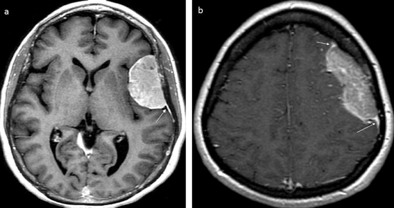
Dural lymphoma (DL) is a rare type of primary CNS lymphoma arising from the dura mater. The optimal treatment is uncertain. A retrospective review was performed on 26 DL patients. Seventeen patients underwent resection and nine had a biopsy. Twenty three patients could be assessed for a response to treatment after surgery. Thirteen received focal radiotherapy (RT), six whole brain RT (WBRT), three chemotherapy alone and one chemotherapy followed by WBRT. Twenty two achieved complete response (CR) and one a partial response (PR). Four patients relapsed (two local and two systemic). Median follow up was 64 months, with median progression free survival (PFS) and OS not reached. Three year PFS was 89% (95% CI 0.64-0.97). All patients are alive at last follow-up, demonstrating that DL is an indolent tumor with long survival. CR is achievable with focal therapy in the majority of cases, but there is a risk for relapses and long-term follow-up is recommended.
Keywords: Dural lymphoma; focal radiotherapy; marginal zone lymphoma; primary central nervous system lymphoma.
Conflict of interest statement
Conflict of interest disclosure statement: The authors have nothing to disclose.
Figures


References
-
- Iwamoto FM, DeAngelis LM, Abrey LE. Primary dural lymphomas: A clinicopathologic study of treatment and outcome in eight patients. Neurology. 2006;66:1763–5. - PubMed
-
- Tu PH, Giannini C, Judkins AR, Schwalb JM, Burack R, O'Neill BP, et al. Clinicopathologic and genetic profile of intracranial marginal zone lymphoma: a primary low-grade CNS lymphoma that mimics meningioma. J Clin Oncol. 2005;23(24):5718–27. - PubMed
-
- Kudrimoti JK, Gaikwad MJ, Puranik SC, Chugh AP. Primary dural non-hodgkin's lymphoma mimicking meningioma: A case report and review of literature. J Cancer Res Ther. 2015;11(3):648. - PubMed
-
- Swerdlow SHCE, Harris NL, Jaffe ES, Pileri SA, Stein H, Thiele J, Vardiman JW. WHO Classification of Tumors of Hematopoietic and Lymphid Tissues. Lyon. 2008
MeSH terms
Grants and funding
LinkOut - more resources
Full Text Sources
Other Literature Sources
Research Materials