Comparative Study
. 2016 Sep;4(18):e12958.
doi: 10.14814/phy2.12958.

Effects of high EPA and high DHA fish oils on changes in signaling associated with protein metabolism induced by hindlimb suspension in rats

Affiliations
Comparative Study

Effects of high EPA and high DHA fish oils on changes in signaling associated with protein metabolism induced by hindlimb suspension in rats

Gabriel Nasri Marzuca-Nassr et al. Physiol Rep. 2016 Sep.

Abstract

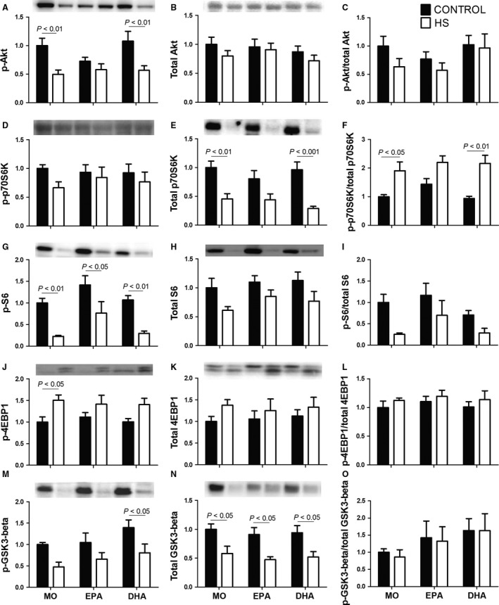
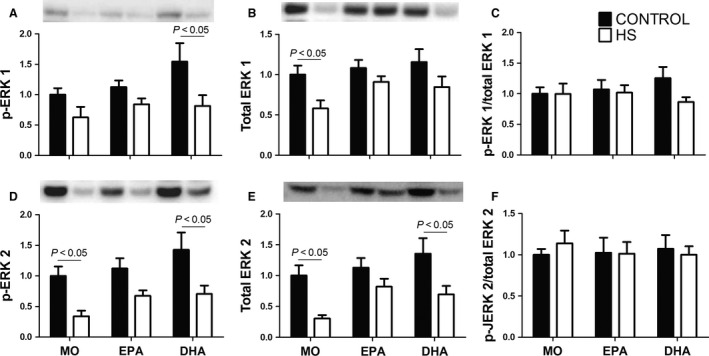
The effects of either eicosapentaenoic (EPA)- or docosahexaenoic (DHA)-rich fish oils on hindlimb suspension (HS)-induced muscle disuse atrophy were compared. Daily oral supplementations (0.3 mL/100 g b.w.) with mineral oil (MO) or high EPA or high DHA fish oils were performed in adult rats. After 2 weeks, the animals were subjected to HS for further 2 weeks. The treatments were maintained alongside HS At the end of 4 weeks, we evaluated: body weight gain, muscle mass and fat depots, composition of fatty acids, cross-sectional areas (CSA) of the soleus muscle and soleus muscle fibers, activities of cathepsin L and 26S proteasome, and content of carbonylated proteins in the soleus muscle. Signaling pathway activities associated with protein synthesis (Akt, p70S6K, S6, 4EBP1, and GSK3-beta) and protein degradation (atrogin-1/MAFbx, and MuRF1) were evaluated. HS decreased muscle mass, CSA of soleus muscle and soleus muscle fibers, and altered signaling associated with protein synthesis (decreased) and protein degradation (increased). The treatment with either fish oil decreased the ratio of omega-6/omega-3 fatty acids and changed protein synthesis-associated signaling. EPA-rich fish oil attenuated the changes induced by HS on 26S proteasome activity, CSA of soleus muscle fibers, and levels of p-Akt, total p70S6K, p-p70S6K/total p70S6K, p-4EBP1, p-GSK3-beta, p-ERK2, and total ERK 1/2 proteins. DHA-rich fish oil attenuated the changes induced by HS on p-4EBP1 and total ERK1 levels. The effects of EPA-rich fish oil on protein synthesis signaling were more pronounced. Both EPA- and DHA-rich fish oils did not impact skeletal muscle mass loss induced by non-inflammatory HS.

Keywords: Hindlimb suspension; muscle atrophy; omega‐3 fatty acids; protein synthesis/degradation.


Figures

Figure 1
Figure 1
Quantitative analysis of western blot membranes stained with Ponceau S. (A) Images of the western blot membranes stained with Ponceau S used in this study. (B) Average quantitative analysis of Ponceau S staining. No significant differences were observed. The results were compared using two‐way ANOVA and Bonferroni post hoc test. MO, Mineral oil supplementation; EPA, High eicosapentaenoic acid fish oil supplementation; DHA, High docosahexaenoic acid fish oil supplementation; HS, hindlimb suspension; MO‐C, Mineral oil supplemented group; MO‐HS, Mineral oil supplemented and hindlimb suspension group; EPA‐C, High eicosapentaenoic acid fish oil supplemented group; EPA‐HS, High eicosapentaenoic acid fish oil supplemented and hindlimb suspension group; DHA‐C, High docosahexaenoic acid fish oil supplemented group; DHA‐HS, High docosahexaenoic acid fish oil supplemented and hindlimb suspension group; P, pool containing a mixture with equal parts of all samples – used to normalize Ponceau S quantitative results; AU, arbitrary units.
Figure 2
Figure 2
Cross‐sectional areas (CSA) of the soleus muscle and soleus muscle fibers. (A) Representative histological hematoxylin and eosin staining images of cross‐sectional areas of the soleus muscle. a. MO‐C; b. MOHS; c. EPA‐C; d. EPAHS; e. DHA‐C; f. DHAHS. Reference bar represents 100 μm. (B) Representative histological hematoxylin and eosin staining images of cross‐sectional areas of soleus muscle fibers; a. MO‐C; b. MOHS; c. EPA‐C; d. EPAHS; e. DHA‐C; f. DHAHS. Reference bar represents 100 μm. (C) Cross‐sectional area of the soleus muscle. Values are presented as mean ± SEM, n = 5–8 animals. The results were compared using two‐way ANOVA and Bonferroni post hoc test. (D) Cross‐sectional areas of soleus muscle fibers. Values are presented as scatter plot with median, n = 750–1199. The results were analyzed as previously described using the 95% CI of the median. (E) Distribution of the soleus muscle fibers according to the ranges of the areas of soleus muscle fibers: 0–2800 μm2. MO‐C, Mineral oil supplemented group; MO‐HS, Mineral oil supplemented and hindlimb suspension group; EPA‐C, High eicosapentaenoic acid fish oil supplemented group; EPA‐HS, High eicosapentaenoic acid fish oil supplemented and hindlimb suspension group; DHA‐C, High docosahexaenoic acid fish oil supplemented group; DHA‐HS, High docosahexaenoic acid fish oil supplemented and hindlimb suspension group; MO, Mineral oil supplementation; EPA, High eicosapentaenoic acid fish oil supplementation; DHA, High docosahexaenoic acid fish oil supplementation; HS, hindlimb suspension. Ω, difference between groups.
Figure 3
Figure 3
(A) The cathepsin L activity in soleus muscle (B) 26S proteasome activity in soleus muscle, (C) content of carbonylated proteins of soleus muscle and (D) representative image of the western blotting analysis of carbonylated proteins (55 KDa‐130 KDa). Values are presented as mean ± SEM on the basis of total protein loading as indicated by the Ponceau S measurement, n = 6–8 animals. The results were compared using two‐way ANOVA and Bonferroni post hoc test. MO, Mineral oil supplementation; EPA, High eicosapentaenoic acid fish oil supplementation; DHA, High docosahexaenoic acid fish oil supplementation; HS, hindlimb suspension; MO‐C, Mineral oil supplemented group; MO‐HS, Mineral oil supplemented and hindlimb suspension group; EPA‐C, High eicosapentaenoic acid fish oil supplemented group; EPA‐HS, High eicosapentaenoic acid fish oil supplemented and hindlimb suspension group; DHA‐C, High docosahexaenoic acid fish oil supplemented group; DHA‐HS, High docosahexaenoic acid fish oil supplemented and hindlimb suspension group; AU, arbitrary units.
Figure 4
Figure 4
Contents of proteins associated with signaling pathway protein synthesis in the soleus muscle: (A) p‐Akt, (B) total Akt, (C) p‐Akt/Akt total ratio, (D) p‐p70S6k, (E) total p70S6k, (F) p‐p70S6k/p70S6k total ratio, (G) p‐S6, (H) total S6, (I) p‐S6/S6 total ratio, (J) p‐4EBP1, (K) total 4EBP1, (L) p‐4EBP1/4EBP1 total ratio, (M) p‐GSK3‐beta, (N) total GSK3‐beta, (O) p‐GSK3‐beta/total GSK3‐beta ratio. Values are presented as mean ± SEM on the basis of total protein loading as indicated by the Ponceau S measurements and expressed relative to MO‐C, n = 6–8 animals. The results were compared using two‐way ANOVA and Bonferroni post hoc test. MO, Mineral oil supplementation; EPA, High eicosapentaenoic acid fish oil supplementation; DHA, High docosahexaenoic acid fish oil supplementation; HS, hindlimb suspension; MO‐C, Mineral oil supplemented group; MO‐HS, Mineral oil supplemented and hindlimb suspension group; EPA‐C, High eicosapentaenoic acid fish oil supplemented group; EPA‐HS, High eicosapentaenoic acid fish oil supplemented and hindlimb suspension group; DHA‐C, High docosahexaenoic acid fish oil supplemented group; DHA‐HS, High docosahexaenoic acid fish oil supplemented and hindlimb suspension group.
Figure 5
Figure 5
Contents of proteins associated with signaling pathway of protein degradation in the soleus muscle: (A) atrogin‐1/MAFbx, (B) MuRF‐1, (C) representative images of the western blotting analysis of atrogin‐1/MAFbx and MuRF‐1. Values are presented as mean ± SEM on the basis of total protein loading as indicated by the Ponceau S measurement and expressed relative to MO‐C, n = 7–8 animals. The results were compared using two‐way ANOVA and Bonferroni post hoc test. MO, Mineral oil supplementation; EPA,High eicosapentaenoic acid fish oil supplementation; DHA, High docosahexaenoic acid fish oil supplementation; HS, hindlimb suspension; MO‐C, Mineral oil supplemented group; MO‐HS, Mineral oil supplemented and hindlimb suspension group; EPA‐C, High eicosapentaenoic acid fish oil supplemented group; EPA‐HS, High eicosapentaenoic acid fish oil supplemented and hindlimb suspension group; DHA‐C, High docosahexaenoic acid fish oil supplemented group; DHA‐HS, High docosahexaenoic acid fish oil supplemented and hindlimb suspension group.
Figure 6
Figure 6
Contents of phosphorylated and total ERK 1 and 2 proteins in the soleus muscle: (A) p‐ERK 1, (B) total ERK 1, (C) p‐ERK 1/total ERK 1 ratio, (D) p‐ERK 2, (E) total ERK 2, (F) p‐ERK 2/total ERK 2 ratio. Values are presented as mean ± SEM on the basis of total protein loading as indicated by the Ponceau S measurements and expressed relative to MO‐C, n = 7–8 animals. The results were compared using two‐way ANOVA and Bonferroni post hoc test. MO, Mineral oil supplementation; EPA, High eicosapentaenoic acid fish oil supplementation; DHA, High docosahexaenoic acid fish oil supplementation; HS, hindlimb suspension; MO‐C, Mineral oil supplemented group; MO‐HS, Mineral oil supplemented and hindlimb suspension group; EPA‐C, High eicosapentaenoic acid fish oil supplemented group; EPA‐HS, High eicosapentaenoic acid fish oil supplemented and hindlimb suspension group; DHA‐C, High docosahexaenoic acid fish oil supplemented group; DHA‐HS, High docosahexaenoic acid fish oil supplemented and hindlimb suspension group.

References

    1. Andrianjafiniony, T. , Dupré‐Aucouturier S., Letexier D., Couchoux H., and Desplanches D.. 2010. Oxidative stress, apoptosis, and proteolysis in skeletal muscle repair after unloading. Am. J. Physiol. Cell Physiol. 299:C307–C315. - PubMed
    1. AOAC . 2005. Official method 996.06. Official methods of analysis of AOAC International, 18th ed. AOAC International, Gaithersburg, MD.
    1. AOCS (2007). Official method Ce 1j‐07. Official methods and recommended practices of the AOCS. 6th ed AOCS, Champaign, IL, USA.
    1. Baehr, L. M. , Furlow J. D., and Bodine S. C.. 2011. Muscle sparing in muscle RING finger 1 null mice: response to synthetic glucocorticoids. J. Physiol. 589:4759–4776. - PMC - PubMed
    1. Baillie, R. A. , Takada R., Nakamura M., and Clarke S. D.. 1999. Coordinate induction of peroxisomal acyl‐CoA oxidase and UCP‐3 by dietary fish oil: a mechanism for decreased body fat deposition. Prostaglandins Leukot. Essent. Fatty Acids 60:351–356. - PubMed

Publication types

MeSH terms

LinkOut - more resources