. 2016 Sep 21:6:33549.
doi: 10.1038/srep33549.

Polyamines release the let-7b-mediated suppression of initiation codon recognition during the protein synthesis of EXT2

Affiliations

Polyamines release the let-7b-mediated suppression of initiation codon recognition during the protein synthesis of EXT2

Masataka Imamura et al. Sci Rep. .

Abstract

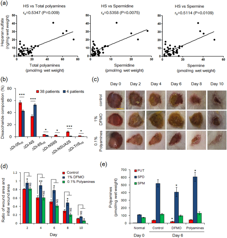
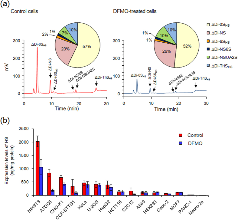
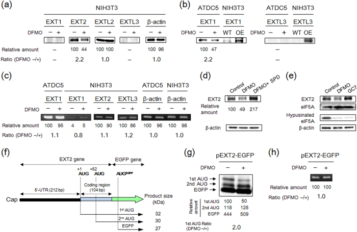
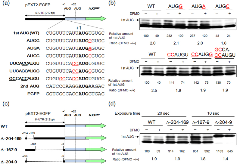
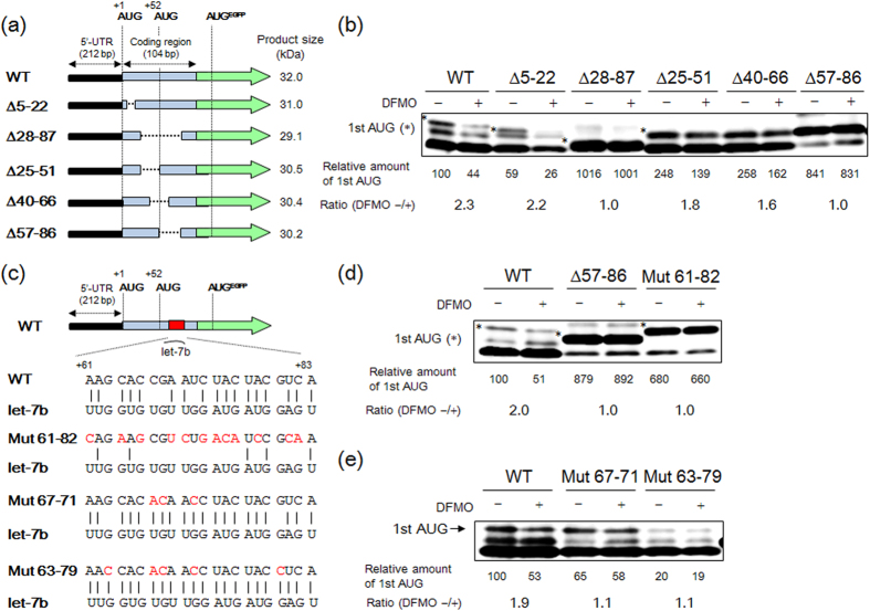
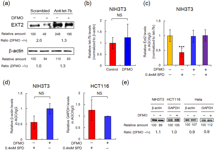
Proteoglycans (PGs), a family of glycosaminoglycan (GAG)-protein glycoconjugates, contribute to animal physiology through interactions between their glycan chains and growth factors, chemokines and adhesion molecules. However, it remains unclear how GAG structures are changed during the aging process. Here, we found that polyamine levels are correlated with the expression level of heparan sulfate (HS) in human skin. In cultured cell lines, the EXT1 and EXT2 enzymes, initiating HS biosynthesis, were stimulated at the translational level by polyamines. Interestingly, the initiation codon recognition by 43S preinitiation complex during EXT2 translation is suppressed by let-7b, a member of the let-7 microRNA family, through binding at the N-terminal amino acid coding sequence in EXT2 mRNA. Let-7b-mediated suppression of initiation codon depends on the length of 5'-UTR of EXT2 mRNA and its suppression is inhibited in the presence of polyamines. These findings provide new insights into the HS biosynthesis related to miRNA and polyamines.


Figures

Figure 1
Figure 1. Relationship between heparan sulfate and polyamines in skin.
(a) Correlation between HS and polyamine contents in human dermis. Median values of HS, total polyamines, spermidine and spermine were 6.2 ng/mg wet weight, 11.3, 3.4 and 7.8 pmol/mg wet weight, respectively. In four subjects increased levels of HS levels were well correlated with total polyamines, spermidine and spermine. Data of 42 subjects with breast reconstruction was evaluated by Spearman’s rank correlation analysis (rs and p value) using GraphPad Prism® Software (GraphPad Software). (b) Disaccharide compositions of unsaturated disaccharides of HS in dermis from four patients having a good correlation between HS and total polyamines and from other 38 patients. Data were evaluated by two-tailed unpaired Student’s t-tests. ⋆⋆⋆P < 0.005, 0.01 < P < 0.05. (c,d) Effect of polyamines on wound healing of mouse skin. Three full thickness wounds were made in individual mouse using an 8-mm biopsy punch. After wounding, hydrophilic petrolatum only (control), hydrophilic petrolatum containing 1% DFMO or 0.1% polyamines (0.075% spermidine, 0.025% spermine) were applied to wounds every day. The scale bar shown is 5 mm. Data are expressed as the mean ± s.e.m. (n = 6). ⋆⋆P < 0.01, 0.01 < P < 0.05, NS, not significant were determined by two-tailed unpaired Student’s t-tests. (e) Determination of polyamine contents in wound regions (surround skin) by HPLC. PUT, putrescine; SPD, spermidine; SPM, spermine. Data are expressed as the mean ± s.e.m. (n = 6). 0.01 < P < 0.05 compared to control skin.
Figure 2
Figure 2. Levels and disaccharide compositions of heparan sulfate in normal and DFMO treated cells.
(a) Chromatogram and disaccharide compositions of HS in NIH3T3 cells cultured with or without DFMO. HS purified from 1 × 107 cells was treated with 1 mIU of heparinase I, II and III, and then submitted to HPLC. Note that the level of HS but not its disaccharide composition clearly changed in DFMO-treated cells. See Supplementary Methods for structures of the disaccharides. (b) Levels of HS in 15 cell types of untreated (control) and DFMO-treated cells. Data are expressed as the mean ± s.e.m. (n = 3). Detailed expression level and disaccharide composition of HS in 15 types of control and DFMO-treated cells are shown in Supplementary Table S9.
Figure 3
Figure 3. EXT1 and EXT2 synthesis are enhanced by polyamines at the level of translation.
(a) Effect of polyamine depletion on the expression level of EXT proteins in NIH3T3 cells. For Western blotting of EXT proteins and β-actin, 30 μg (EXT1), 20 μg (EXT2), 20 μg (EXTL2), 60 μg (EXTL3) or 5 μg (β-actin) of protein of whole cell lysate, prepared from cells cultured with or without DFMO, was used. (b) Effect of polyamine depletion on the expression of EXT1 and EXTL3 in ATDC5 cells. For Western blotting, 30 μg (EXT1) or 60 μg (EXTL3) of protein was used. OE: over expressed. (c) Effect of polyamine depletion on the expression level of mRNAs of EXT gene family in NIH3T3 and ATDC5 cells. (d) Effects of 5 mM DFMO and/or 25 μM spermidine (SPD) on the expression level of EXT2 in NIH3T3 cells. To avoid the degradation of SPD, 1 mM aminoguanidine, an inhibitor of serum amine oxidase, was added together with SPD in culture medium for 3 days. (e) Effect of GC7, an efficient inhibitor of deoxyhypusin synthase, on the expression of EXT2 protein in NIH3T3 cells. GC7 (20 μM) was added to culture medium for 3 days to inhibit the hypusination for eIF5A. Note that the EXT2 expression was maintained despite an inhibition of hypusination of eIF5A by the GC7 treatment. For detection of the eIF5A protein, 10 μg of protein was used. (f) Structure of EXT2-EGFP fusion genes. Two kinds of fusion proteins (32 kDa and 30 kDa) and EGFP protein (27 kDa) can be produced by individual AUG triplets in EXT2-EGFP fusion gene. (g) Effect of polyamine depletion on the expression level of EXT2-EGFP fusion proteins in NIH3T3 cells. For detection of the EXT2-EGFP fusion protein, 30 μg of protein was used. (h) Effect of polyamine depletion on the expression level of EXT2-EGFP mRNA. Experiments were repeated in triplicate with reproducible results.
Figure 4
Figure 4. Effect of AUG codon context and 5′-UTR on the polyamine stimulation of EXT2 synthesis.
(a) Structures of mutated AUG codon context in EXT2-EGFP fusion genes. The modified nucleotides in first AUG context are shown in red and underlined. (b) Effect of mutated first AUG codon context in EXT2 gene on the expression level of the first AUG product of the EXT2-fusion protein in NIH3T3 cells. (c) Structures of 5′-UTR deletion mutants of EXT2-EGFP fusion genes. (d) Effect of 5′-UTR on the expression level of first AUG product of EXT2-fusion protein in NIH3T3 cells. In the case of the 5′-UTR deletion mutants, exposure time of ECL detection was shortened compared to the WT because of a significant increase in the expression of EGFP-protein. Transfection and DFMO treatment were carried out as described under “Methods” and 30 μg of protein was used to detect the EGFP fusion protein. Experiments were repeated in triplicate with reproducible results.
Figure 5
Figure 5. Effect of let-7b binding site in N-terminal amino acid coding sequence on polyamine stimulation of EXT2 synthesis.
(a) Structures of deletion mutants of N-terminal CDS in EXT2-EGFP fusion genes. Based on the possible secondary structure of EXT2 mRNA obtained by Mfold (http://unafold.rna.albany.edu/), specified region of CDS was deleted (See Supplementary Fig. S8). Note that sizes of first AUG products in deletion mutants were smaller than that of WT. (b) Effect of N-terminal CDS on the expression levels of first AUG products (*) of EXT2-EGFP fusion proteins in NIH3T3 cells. (c) Sequence of let-7b binding site in EXT2-EGFP fusion gene. (d,e) Effects of mutations of let-7b binding site on the expression level of the first AUG products of the EXT2-EGFP fusion proteins in NIH3T3 cells. Molecular weight of the first AUG product from ∆57-86 mutant is 30.2 kDa. For detection of the EGFP fusion protein, 30 μg of protein was used. Experiments were repeated in triplicate with reproducible results.
Figure 6
Figure 6. Direct inhibition of let-7b binding by spermidine to its target sequence.
(a) Effect of anti-let-7b on the expression level of EXT2 protein in NIH3T3 cells. The microRNA inhibitor for let-7b or negative control A (scrambled) were transfected to NIH3T3 cells for 72 h in the presence or absence of 5 mM DFMO. (b) Effect of polyamine depletion on the expression level of let-7b in NIH3T3 cells was examined by qPCR. The level of let-7b expression in control cells was defined as 1.0. NS, not significant, two-tailed unpaired Student’s t-tests. (c,d) Immunoprecipitation (IP) of miRNA/EXT2 mRNA (c), β-actin or GAPDH mRNAs (d) complexes associated with Argonate proteins (Ago1, Ago2 or Ago3) from NIH3T3 or HCT116 cell extract. Precipitated miRNA/mRNA complexes were treated with proteinase K, and resulting free mRNAs were converted to cDNA and quantified by qPCR. The results obtained with Ago-IP were normalized by mouse IgG-IP as a negative control. (c) In this pull-down assay system, since precipitated let-7b by Ago-IP was also detectable, EXT2 mRNA was further normalized to let-7b. Data are expressed as the mean ± s.e.m. of three independent experiments and data was by one-way ANOVA with Tukey-Kramer post-test using GraphPad Prism® Software (GraphPad Software). ⋆⋆⋆P < 0.005. (d) In case of values of β-actin and GAPDH, both of which are obtained from three independent experiments are expressed as the mean ± s.e.m. Data were evaluated by two-tailed unpaired Student’s t-tests, however no significant difference was observed. (e) Effect of polyamine depletion on the expression of β-actin and GAPDH.

Similar articles

Cited by

References

    1. Roden L. Structure and metabolism of connective tissue proteoglycans. 267–371 (Plenum press, New York, 1980).
    1. Bishop J. R., Schuksz M. & Esko J. D. Heparan sulphate proteoglycans fine-tune mammalian physiology. Nature 446, 1030–1037, doi: 10.1038/nature05817 (2007). - DOI - PubMed
    1. Mikami T. & Kitagawa H. Biosynthesis and function of chondroitin sulfate. Biochim Biophys Acta 1830, 4719–4733, doi: 10.1016/j.bbagen.2013.06.006 (2013). - DOI - PubMed
    1. Maeda N. Proteoglycans and neuronal migration in the cerebral cortex during development and disease. Front Neurosci 9, 98, doi: 10.3389/fnins.2015.00098 (2015). - DOI - PMC - PubMed
    1. Sarrazin S., Lamanna W. C. & Esko J. D. Heparan sulfate proteoglycans. Cold Spring Harb Perspect Biol 3, doi: 10.1101/cshperspect.a004952 (2011). - DOI - PMC - PubMed

Publication types

MeSH terms

LinkOut - more resources