. 2016 Sep 21:7:12861.
doi: 10.1038/ncomms12861.

A conserved influenza A virus nucleoprotein code controls specific viral genome packaging

Affiliations

A conserved influenza A virus nucleoprotein code controls specific viral genome packaging

Étori Aguiar Moreira et al. Nat Commun. .

Abstract

Packaging of the eight genomic RNA segments of influenza A viruses (IAV) into viral particles is coordinated by segment-specific packaging sequences. How the packaging signals regulate the specific incorporation of each RNA segment into virions and whether other viral or host factors are involved in this process is unknown. Here, we show that distinct amino acids of the viral nucleoprotein (NP) are required for packaging of specific RNA segments. This was determined by studying the NP of a bat influenza A-like virus, HL17NL10, in the context of a conventional IAV (SC35M). Replacement of conserved SC35M NP residues by those of HL17NL10 NP resulted in RNA packaging defective IAV. Surprisingly, substitution of these conserved SC35M amino acids with HL17NL10 NP residues led to IAV with altered packaging efficiencies for specific subsets of RNA segments. This suggests that NP harbours an amino acid code that dictates genome packaging into infectious virions.


Figures
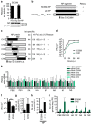
Figure 1
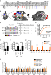
Figure 1. Limited compatibility of SC35M/bat NP chimeras to rescue and package viral genomes of SC35M.
(a) SC35M polymerase activity in the presence of Bat NP. HEK293T cells were transiently transfected with expression plasmids coding for PB2, PB1, PA of SC35M, the indicated NP proteins, a minigenome encoding the firefly luciferase and a Renilla luciferase expression plasmid to normalize for variations in transfection efficiency. In the negative control (Neg.) PB1 was omitted. Western blot analysis was performed to determine the expression levels of NP. (b) Cartoon depicting NP segments of A/SC35M (SC35M NP), HL17NL10 (Bat NP) or a NP segment (SC35M250-NPORF-Bat) harbouring the non-coding regions of SC35M NP, 5′ and 3′ coding sequences of SC35M NP and the complete ORF of Bat NP. +successful rescue; −no rescue. (c) Relative SC35M polymerase activities in the presence of the mutant NP proteins (CH1–CH5). Mean and s.d. of three independent experiments are indicated in parenthesis. SC35M rescue experiments were performed with NP segments encoding the indicated mutant proteins. +successful rescue; −no rescue. (d) MDCKII cells were infected at an MOI of 0.001 with wt SC35M or rCH2. At the indicated time points post infection (p.i.), virus titres were determined by plaque assay. (e) Relative ratio of the viral transcript level in wt SC35M- or rCH2-infected cells. Steady state levels of viral transcripts (mRNA, cRNA and vRNA) and 5S ribosomal RNA (5S rRNA) were determined by primer extension analysis using total RNA from MDCKII cells infected at an MOI of 5 with wt SC35M or rCH2 for 6 h. Signal intensities were normalized to the signal intensities obtained with 5S rRNA. Normalized values obtained in wt SC35M-infected cells were set to 1 (all non-significant). (f) Viral infectivity of SC35M and rCH2 (PFU) using identical HA titre. **P<0.01. (g) Relative ratio of the number of viral particles (counted by electron microscopy) divided by the number of infectious particles (determined by plaque assay) between wt SC35M and rCH2. Values obtained for SC35M were set to 1. *P<0.05. (h) Ratio of incorporated NP protein in viral particles between SC35M and rCH2. Protein levels were determined by Western blot analysis of virus stocks with equal infectivity (PFU). **P<0.01. (i) Relative ratio of genome segments in viral particles preparations of SC35M and rCH2. RNA was prepared from virus stocks with equal PFU and subjected to quantitative RT-PCR. Levels of viral genome transcripts obtained with SC35M were set to 1. *P<0.05; **P<0.01; ***P<0.001. Student's t test was used for two-group comparisons. Error bars indicate the mean and s.d. of at least three independent experiments.
Figure 2
Figure 2. Substitution of 14 Bat NP-specific amino acids in CH4 is required to rescue SC35M.
(a) Alignment of SC35M NP and CH4 variants. Bat NP-specific amino acids are indicated in red. (b) The 19 Bat NP-specific amino acids in CH4 (highlighted in black) were mutated to SC35M-specific amino acids leaving either 14 (CH4.14), 4 (CH4.4) or no Bat NP-specific amino acids (CH4.0) and 100, 86 and 80 Bat NP-specific nucleotides, respectively. Relative polymerase activities of the indicated CH4 variants were determined by polymerase reconstitution assays in HEK293T cells. Mean and s.d. of three independent experiments are indicated in parenthesis. +successful rescue; −no rescue of recombinant viruses. (c) MDCKII cells were infected at an MOI of 0.001 with either wt SC35M, rCH4.4 or SC35M-CH4.0. At the indicated time points post infection (p.i.) virus titres were determined by plaque assay. Error bars indicate the mean and s.d. of at least three independent experiments.
Figure 3
Figure 3. A subset of Bat NP-specific amino acids of CH4 causes severe attenuation and irregular packaging of genome segments.
(a) Alignment of SC35M NP and NP mutant proteins harbouring 14 (NP14), 10 (NP10) or 7 (NP7) Bat NP-specific amino acids of CH4. Upper panel shows the sequence logo of the consensus sequence of NP of available IAV strains (n=27,675 strains). (b) Positions of Bat NP-specific amino acids in the modelled crystal structure of SC35M NP. The program PyMOL was used to assign the indicated positions. Bat-specific amino acids are marked in the colour code used in a. Note that amino acid 239 is not surface exposed. (c) Relative SC35M polymerase activities in the presence of the mutant NP proteins determined by polymerase reconstitution assays. Bat-specific amino acids and nucleotides are indicated. SC35M rescue experiments were performed with NP segments encoding the indicated mutant proteins. +successful rescue; −no rescue. (d) MDCKII cells were infected at an MOI of 0.001 with wt SC35M or recombinant virus rNP7. At the indicated time post infection (p.i.), virus titres were determined by plaque assay. (e) Relative ratio of the number of viral particles (counted by electron microscopy) divided by the number of infectious particles (determined by plaque assay) between wt SC35M and rNP7. Values obtained for SC35M were set to 1. *P<0.05. (f) Ratio of incorporated NP and M1 proteins in viral particles between SC35M and rNP7. Protein levels were determined by Western blot analysis of virus stocks with equal infectivity (PFU). *P<0.05; ***P<0.001. (g) Relative ratio of genome segments identified in viral particles preparations of SC35M and rNP7. RNA was prepared from virus stocks with identical infectivity (PFU) and subjected to quantitative RT-PCR. Levels of viral genome transcripts obtained with wt SC35M were set to 1. *P<0.05; **P<0.01; ***P<0.001. (h) Relative ratio of the viral transcript level in wt SC35M- or rNP7-infected cells. Steady state levels of viral transcripts (mRNA, cRNA and vRNA) and 5S ribosomal RNA (5S rRNA) were determined by primer extension analysis using total RNA from MDCKII cells infected at an MOI of 5 with wt SC35M or rCH2 for 6 h. Signal intensities were normalized to the signal intensities obtained with 5S RNA. Normalized values obtained in cells infected with wt SC35M were set to 1. *P<0.05; **P<0.01; ***P<0.001. Student's t test was used for two-group comparisons. Error bars indicate the mean and s.d. of three independent experiments.
Figure 4
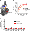
Figure 4. The compensatory amino acid substitution R31G in NP7 improves viral growth and genome packaging.
(a) Positions of Bat NP-specific amino acids in the head domain and R31G body domain of NP7 using the modelled crystal structure of SC35M NP. The program PyMOL was used to assign the indicated positions. Bat-specific amino acids are marked in the colour code used in Fig. 3b. (b) MDCKII cells were infected at an MOI of 0.001 with wt SC35M, rNP7 or rNP7-R31G. At the indicated time post infection (p.i.), viral titres were determined by plaque assay. *P<0.05; **P<0.01; ****P<0.0001. (c) Relative ratio of genome segments identified in viral particles preparations of SC35M and rNP7-R31G. RNA was prepared from virus stocks with identical infectivity (PFU) and subjected to quantitative RT-PCR. Levels of viral genome transcripts obtained with wt SC35M were set to 1. *P<0.05; **P<0.01. Student's t test was used for two-group comparisons. Error bars indicate the mean and s.d. of at least three independent experiments.
Figure 5
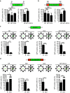
Figure 5. Bat NP-specific amino acids in SC35M NP cause impaired packaging.
(a,b) Packaging efficiency of a single minigenome supported by SC35M NP (NP), CH2, NP7 or NP7-R31G in a VLP-based packaging assay. Reconstitutions of SC35M VLPs were carried out in the presence a GFP-encoding reporter segment flanked by IS of the NP segment (IS−GFP) or in addition with the bundling signals (BS) in the 5′ and 3′ NP ORF (IS+BS−GFP). After infection of MDCKII cells and subsequent superinfection with wt SC35M, GFP-positive cells were quantified by FACS. NS, not significant; *P<0.05; **P<0.01. (c,d) Packaging efficiency mediated by SC35M NP (NP), NP7, CH2 or NP7-R31G in the presence of the reporter segment IS+GFP (R) (c) or IS+BS−GFP (R) (d) and, if indicated, in the presence of the remaining seven wild-type genome segments (+7). NS, not significant; *P<0.05; **P<0.01; ****P<0.0001. Student's t test was used for two-group comparisons. Error bars indicate the mean and s.d. of at least three independent experiments.
Figure 6
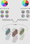
Figure 6. Genome packaging sequence or NP code mutations disrupt coordinated packaging of eight influenza genome segments into viral particles.
Wt NP code and vRNA packaging sequences ensure coordinated incorporation of the eight different genome segments into influenza A virus particles, resulting in an equal ratio of the individual viral genome segments (left panel). As indicated by dashed lines, packaging could be coordinated by various interactions between segment-specific vRNPs, including interactions between vRNA packaging sequences, between vRNA packaging sequences in one vRNP and amino acids of the NP code (indicated in yellow) in a second vRNP, and direct interactions of the NP code amino acids. Mutations in the NP code and/or vRNA packaging sequence result in the loss of coordinated packaging of the eight different genome segments into viral particles and a disproportional ratio of the viral segments (right panel). Loss of coordinated packaging might be caused by impaired interactions between vRNPs mediated by amino acids of the NP code and/or nucleotides of the RNA packaging sequences.

Similar articles

Cited by

References

    1. Eisfeld A. J., Neumann G. & Kawaoka Y. At the centre: influenza A virus ribonucleoproteins. Nat. Rev. Microbiol. 13, 28–41 (2015). - PMC - PubMed
    1. Zheng W. & Tao Y. J. Structure and assembly of the influenza A virus ribonucleoprotein complex. FEBS Lett. 587, 1206–1214 (2013). - PubMed
    1. Webster R. G., Bean W. J., Gorman O. T., Chambers T. M. & Kawaoka Y. Evolution and ecology of influenza A viruses. Microbiol. Rev. 56, 152–179 (1992). - PMC - PubMed
    1. Lu L., Lycett S. J. & Leigh Brown A. J. Reassortment patterns of avian influenza virus internal segments among different subtypes. BMC Evol. Biol. 14, 16 (2014). - PMC - PubMed
    1. Steel J. & Lowen A. C. Influenza A virus reassortment. Curr. Top. Microbiol. Immunol. 385, 377–401 (2014). - PubMed

Publication types