. 2016 Oct 25;7(43):69976-69990.
doi: 10.18632/oncotarget.12099.

Fibroblast growth factor receptor 4 induced resistance to radiation therapy in colorectal cancer

Affiliations

Fibroblast growth factor receptor 4 induced resistance to radiation therapy in colorectal cancer

Mohamed A Ahmed et al. Oncotarget. .

Erratum in

Abstract

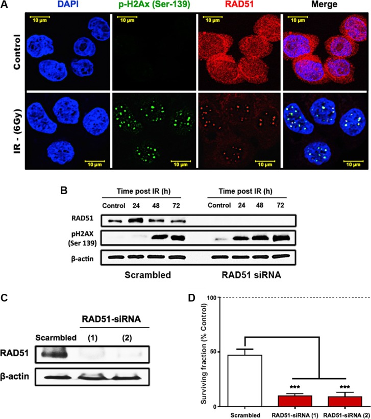
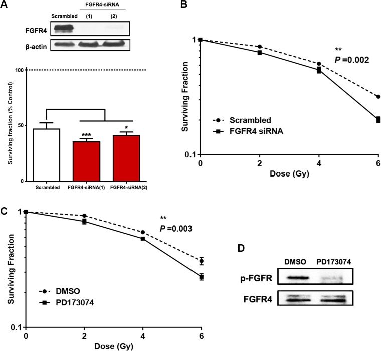
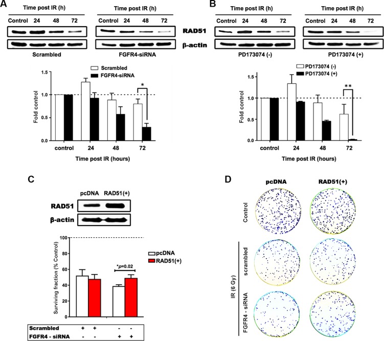
In colorectal cancer (CRC), fibroblast growth factor receptor 4 (FGFR4) is upregulated and acts as an oncogene. This study investigated the impact of this receptor on the response to neoadjuvant radiotherapy by analyzing its levels in rectal tumors of patients with different responses to the therapy. Cellular mechanisms of FGFR4-induced radioresistance were analyzed by silencing or over-expressing FGFR4 in CRC cell line models. Our findings showed that the FGFR4 staining score was significantly higher in pre-treatment biopsies of non-responsive than responsive patients. Similarly, high expression of FGFR4 inhibited radiation response in cell line models. Silencing or inhibition of FGFR4 resulted in a reduction of RAD51 levels and decreased survival in radioresistant HT29 cells. Increased RAD51 expression rescued cells in the siFGFR4-group. In radiosensitive SW480 and DLD1 cells, enforced expression of FGFR4 stabilized RAD51 protein levels resulting in enhanced clearance of γ-H2AX foci and increased cell survival in the mismatch repair (MMR)-proficient SW480 cells. MMR-deficient DLD1 cells are defective in homologous recombination repair and no FGFR4-induced radioresistance was observed. Based on our results, FGFR4 may serve as a predictive marker to select CRC patients with MMR-proficient tumors who may benefit from pre-operative radiotherapy.

Keywords: FGFR4; RAD51; colorectal cancer; radiotherapy.


Conflict of interest statement

CONFLICTS OF INTEREST

The authors declare no potential conflicts of interest.

Figures

Figure 1
Figure 1. Distribution of staining-based FGFR4 and RAD51 expression in pre-neoadjuvant rectal cancer biopsies
Representative staining of biopsies exhibiting negative, weak, moderate and strong FGFR4 (A) or RAD51 (B) staining. Rectal cancer tissues were classified according to overall staining intensity for FGFR4 (C) and RAD51 (D), based on slide scans and morphometric analysis. Scale bar = 50 μm.
Figure 2
Figure 2. FGFR4 correlates with RAD51 protein levels and poor clinical outcome in human rectal cancer
FGFR4 (A) and RAD51 (B) staining intensity in pre-treatment biopsies was scored for responders and non-responders, according to the immunoreactive scoring (IRS) described in the “materials and methods.” The figures show the individual values together with the mean intensity score ± SEM, *p < 0.05 − t-test. Representative staining of FGFR4 (C) and RAD51 (D) in a resected rectal tumor of a patient who did not respond to the neoadjuvant chemoradiotherapy regimen. Scale bar = 100 μm.
Figure 3
Figure 3. FGFR4 expression is upregulated after irradiation in a dose-dependent manner together with key homologous recombination-related proteins
Expression of (A) FGFR4, (B) RAD51, (C) BRCA1 and (D) BRCA2 genes were determined by qPCR, 24 h after exposure to different doses of γ-radiation (0, 2, 4 and 6 Gy) in HT29 cells. The expression levels were calculated relative to GAPDH. (E) Cell cycle distribution of HT29 cells irradiated with a single 6 Gy dose of γ-rays. Analysis was done using FACS at 24, 48 and 72 hours post irradiation. (F) Western blot of cdc2 phosphorylation (Tyr-15) status, Cyclin B1 expression, and the histone H3 phosphorylation (Ser-10) in HT29 cells at different time points after exposure to 6 Gy dose. Beta actin was used as loading control.
Figure 4
Figure 4. RAD51-dependent HR is a crucial mediator of HT29 survival after irradiation
(A) Immunofluorescence of RAD51 and γ-H2AX foci formation post IR. HT29 cells were seeded onto cover slips and treated with a single 6 Gy dose of γ-rays. 24 h after IR, cells were formalin fixed, permeabilized, and stained with RAD51 and γ-H2AX antibodies. (B) Western blots showing the effect of RAD51 knockdown on the damage marker, γ-H2AX. (C) Western blots confirming the efficiency of the two tested RAD51 siRNAs. (D) Clonogenic surviving fractions of scrambled/RAD51 siRNA treated HT29 cells showing increased cell killing and induced radiosensitivity of the radioresistant HT29 cells after RAD51 knockdown. Cells were exposed to a single 6 Gy dose of γ-rays, and the surviving fractions were calculated by dividing the number of colonies counted by the corresponding number of cells seeded as described in “Materials and Methods.”
Figure 5
Figure 5. Silencing of FGFR4 induced loss of survival in radioresistant HT29 cells
The knockdown of FGFR4 expression in HT29 cells was achieved using siRNA targeting FGFR4 one day before exposure to a single 6 Gy dose of γ-rays. Two different FGFR4 siRNAs were used and the knockdown efficiency was confirmed by western blot (A, upper panel) and qRT-PCR (A, lower panel). (B) Colony formation assay showed a significant decrease in the surviving fraction of FGFR4-knocked down HT29 cells. (C) Exposure of the cells to the FGFR-inhibitor PD173074 3 hours before exposure to a single 6 Gy dose caused a similar decrease in colony formation capacity of HT29 cells. HT29 cultures were treated with 2 μM PD173074 to inhibit FGFR4-dependent signaling. The efficacy of the inhibitor in blocking the phosphorylation and the activation of the receptor was confirmed with western blot (D).
Figure 6
Figure 6. FGFR4-mediated radiation response involves regulation of RAD51
Proteins were isolated at the indicated time points after irradiation from cultures treated with siRNA targeting FGFR4 (A) or with the FGFR-inhibitor PD173074 (B). The upper panels show typical western blots of RAD51 protein expression in irradiated HT29 cells. The lower panels depict the quantification of RAD51 protein expression from 3 independent experiments normalized to control. (C) RAD51 was overexpressed using a RAD51 expressing vector in HT29 cells as confirmed by western blotting (upper panel). HT29 cells seeded into 6 well plates were transiently transfected with a vector expressing RAD51 or the control vector. For determination of radiation response, cells were co-transfected with RAD51 or control vectors together with either scrambled or FGFR4 siRNAs, before exposure to a single 6 Gy dose of γ-rays. The surviving fraction of the transfected cells was measured by quantification of colonies (lower panel). (D) Representative images of the clonogenicity assay.
Figure 7
Figure 7. FGFR4 silencing-induced damage persistence in HT29 cells
Immunofluorescence of γ-H2AX foci post IR was performed as described in Materials and Methods, and Figure 4. After FGFR4 knockdown (A) or PD173074 treatment (B), cells were treated with a single 6 Gy dose of γ-rays and stained for γ-H2AX 24 h later. Upper panels show representative photographs, lower panels show the quantification of γ-H2AX-foci.
Figure 8
Figure 8. FGFR4 overexpression induced radioresistance
FGFR4 was overexpressed in MMR-proficient SW480 cells and in MMR-deficient DLD1 cells and overexpression was verified by western blotting. After γ-irradiation the surviving cell fraction was increased in the FGFR4-SW480 cells (A), but not in the FGFR4-DLD1 cells (B). Western blot showing stabilization of RAD51 protein levels of irradiated FGFR4 overexpressing SW480 (C) and DLD1 (D) cells, as compared to pcDNA-transfected cells. RAD51 protein lysates were collected at 24, 48 and 72 h after single 6 Gy dose. Fluorescence staining of γ-H2AX in formalin fixed SW480 (E) and DLD1 (F) cells 24 h after irradiation with a single 6 Gy dose. Upper panels show representative pictures of γ-H2AX foci (green fluorescence), lower panels show the quantification of γ-H2AX foci relative to control. Quantification of the HR capacity in SW480 (G) and DLD1 (H) cells, represented by the ratio of GFP+ cells to DsRed+ cells, as described in the Matrials and Methods. The bars represent the mean of 3 independent cultures ± SEM.

Similar articles

Cited by

References

    1. Ramzan Z, Nassri AB, Huerta S. Genotypic characteristics of resistant tumors to pre-operative ionizing radiation in rectal cancer. World J Gastrointest Oncol. 2014;6:194–210. - PMC - PubMed
    1. Curtin NJ. DNA repair dysregulation from cancer driver to therapeutic target. Nat Rev Cancer. 2012;12:801–17. - PubMed
    1. Kuo LJ, Yang LX. Gamma-H2AX - a novel biomarker for DNA double-strand breaks. In Vivo (Brooklyn) 2008;22:305–9. - PubMed
    1. Tarsounas M, Davies AA, West SC. RAD51 localization and activation following DNA damage. Philos Trans R Soc Lond B Biol Sci. 2004;359:87–93. - PMC - PubMed
    1. Vispé S, Cazaux C, Lesca C, Defais M. Overexpression of Rad51 protein stimulates homologous recombination and increases resistance of mammalian cells to ionizing radiation. Nucleic Acids Res. 1998;26:2859–64. - PMC - PubMed

Substances