

Baseline Fourier-Domain Optical Coherence Tomography Structural Risk Factors for Visual Field Progression in the Advanced Imaging for Glaucoma Study
- PMID: 27651070
- PMCID: PMC5121039
- DOI: 10.1016/j.ajo.2016.09.015
Baseline Fourier-Domain Optical Coherence Tomography Structural Risk Factors for Visual Field Progression in the Advanced Imaging for Glaucoma Study
Abstract
Purpose: To identify baseline structural parameters that predict the progression of visual field (VF) loss in patients with open-angle glaucoma.
Design: Multicenter cohort study.
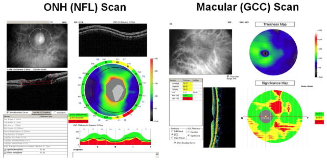
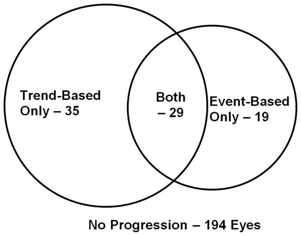
Methods: Participants from the Advanced Imaging for Glaucoma (AIG) study were enrolled and followed up. VF progression is defined as either a confirmed progression event on Humphrey Progression Analysis or a significant (P < .05) negative slope for VF index (VFI). Fourier-domain optical coherence tomography (FDOCT) was used to measure optic disc, peripapillary retinal nerve fiber layer (NFL), and macular ganglion cell complex (GCC) thickness parameters.
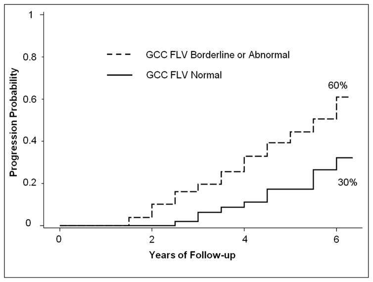
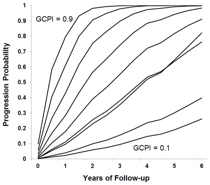
Results: A total of 277 eyes of 188 participants were followed up for 3.7 ± 2.1 years. VF progression was observed in 83 eyes (30%). Several baseline NFL and GCC parameters, but not disc parameters, were found to be significant predictors of progression on univariate Cox regression analysis. The most accurate single predictors were the GCC focal loss volume (FLV), followed closely by NFL-FLV. An abnormal GCC-FLV at baseline increased risk of progression by a hazard ratio of 3.1. Multivariate Cox analysis showed that combining age and central corneal thickness with GCC-FLV in a composite index called "Glaucoma Composite Progression Index" (GCPI) further improved the accuracy of progression prediction. GCC-FLV and GCPI were both found to be significantly correlated with the annual rate of change in VFI.
Conclusion: Focal GCC and NFL loss as measured by FDOCT are the strongest predictors for VF progression among the measurements considered. Older age and thinner central corneal thickness can enhance the predictive power using the composite risk model.
Copyright © 2016 Elsevier Inc. All rights reserved.
Figures
References
-
- Quigley HA, Enger C, Katz J, et al. Risk factors for the development of glaucomatous visual field loss in ocular hypertension. Arch Ophthalmol. 1994;112:644–649. - PubMed
-
- The Advanced Glaucoma Intervention Study (AGIS): 7. The relationship between control of intraocular pressure and visual field deterioration.The AGIS Investigators. Am J Ophthalmol. 2000;130(4):429–440. - PubMed
-
- Leske MC, Heijl A, Hussein M, Bengtsson B, Hyman L, Komaroff E. Factors for glaucoma progression and the effect of treatment: the early manifest glaucoma trial. Arch Ophthalmol. 2003;121(1):48–56. - PubMed
-
- Nouri-Mahdavi K, Hoffman D, Coleman AL, et al. Predictive factors for glaucomatous visual field progression in the Advanced Glaucoma Intervention Study. Ophthalmology. 2004;111(9):1627–1635. - PubMed
-
- Caprioli J, Coleman AL. Intraocular pressure fluctuation a risk factor for visual field progression at low intraocular pressures in the advanced glaucoma intervention study. Ophthalmology. 2008;115(7):1123–1129. e1123. - PubMed
Publication types
MeSH terms
Grants and funding
LinkOut - more resources
Full Text Sources
Other Literature Sources
Medical
