

Interleaving cerebral CT perfusion with neck CT angiography. Part II: clinical implementation and image quality
- PMID: 27651144
- PMCID: PMC5408041
- DOI: 10.1007/s00330-016-4592-z
Interleaving cerebral CT perfusion with neck CT angiography. Part II: clinical implementation and image quality
Abstract
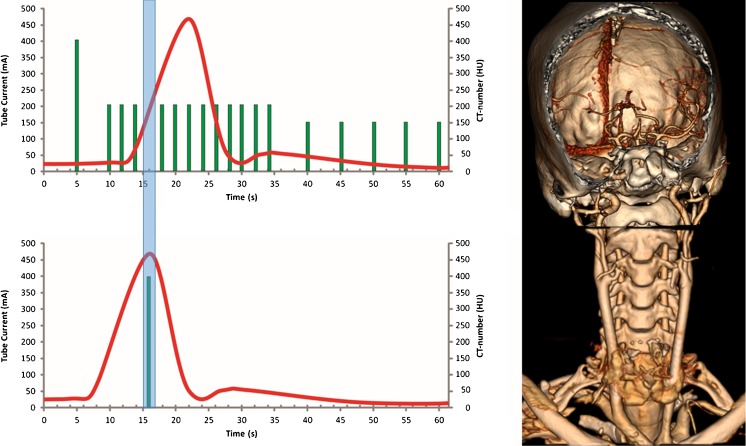
Objectives: Feasibility evaluation of the One-Step Stroke Protocol, which is an interleaved cerebral computed tomography perfusion (CTP) and neck volumetric computed tomography angiography (vCTA) scanning technique using wide-detector computed tomography, and to assess the image quality of vCTA.
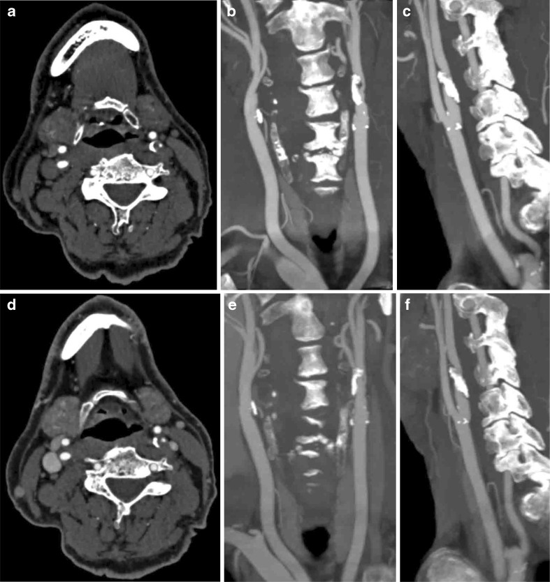
Methods: Twenty patients with suspicion of acute ischaemic stroke were prospectively scanned and evaluated with a head and neck CTA and with the One-Step Stroke Protocol. Arterial enhancement and contrast-to-noise ratio (CNR) in the carotid arteries was assessed. Three observers scored artefacts and image quality of the cervical arteries. The total z-coverage was evaluated.
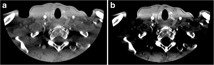
Results: Mean enhancement in the carotid bifurcation was rated higher in the vCTA (595 ± 164 HU) than CTA (441 ± 117 HU). CNR was rated higher in vCTA. Image quality scores showed no significant difference in the region of the carotid bifurcation between vCTA and CTA. Lower neck image quality scores were slightly lower for vCTA due to artefacts, although not rated as diagnostically relevant. In ten patients, the origin of the left common carotid artery was missed by 1.6 ± 0.8 cm. Mean patient height was 1.8 ± 0.09 m. Carotid bifurcation and origin of vertebral arteries were covered in all patients.
Conclusions: The One-Step Stroke Protocol is feasible with good diagnostic image quality of vCTA, although full z-coverage is limited in tall patients.
Key points: • Interleaving cerebral CTP with neck CTA (One-Step Stroke Protocol) is feasible • Diagnostic quality of One-Step Stroke Protocol neck CTA is similar to conventional CTA • One-Step Stroke Protocol neck CTA suffers from streak artefacts in the lower neck • A limitation of One-Step Stroke Protocol CTA is lack of coverage in tall patients • Precise planning of One-Step Stroke Protocol neck CTA is necessary in tall patients.
Keywords: Angiography; Brain; Multidetector computed tomography; Perfusion; Stroke.
Figures
Similar articles
-
Single rotation CTA of extracranial carotids integrated with cerebral CTP provides sufficient quality for decision making in patients with ischaemic stroke.Neuroradiol J. 2021 Apr;34(2):105-112. doi: 10.1177/1971400920974584. Epub 2020 Dec 2. Neuroradiol J. 2021. PMID: 33263488 Free PMC article.
-
Interleaving cerebral CT perfusion with neck CT angiography part I. Proof of concept and accuracy of cerebral perfusion values.Eur Radiol. 2017 Jun;27(6):2649-2656. doi: 10.1007/s00330-016-4577-y. Epub 2016 Oct 7. Eur Radiol. 2017. PMID: 27718078 Free PMC article.
-
Evaluation of virtual monoenergetic imaging algorithms for dual-energy carotid and intracerebral CT angiography: Effects on image quality, artefacts and diagnostic performance for the detection of stenosis.Eur J Radiol. 2018 Feb;99:111-117. doi: 10.1016/j.ejrad.2017.12.024. Epub 2017 Dec 30. Eur J Radiol. 2018. PMID: 29362140
-
Brain perfusion CT in acute stroke: current status.Eur J Radiol. 2003 Mar;45 Suppl 1:S11-22. doi: 10.1016/s0720-048x(02)00359-5. Eur J Radiol. 2003. PMID: 12598022 Review.
-
Systematic Review of CT Angiography in Guiding Management in Pediatric Oropharyngeal Trauma.Laryngoscope. 2023 Mar;133(3):457-466. doi: 10.1002/lary.30179. Epub 2022 May 13. Laryngoscope. 2023. PMID: 35561004
Cited by
-
Robust Segmentation of the Full Cerebral Vasculature in 4D CT of Suspected Stroke Patients.Sci Rep. 2017 Nov 15;7(1):15622. doi: 10.1038/s41598-017-15617-w. Sci Rep. 2017. PMID: 29142240 Free PMC article.
-
A "one-stop-shop" 4D CTA protocol using 320-row CT for advanced imaging in acute ischemic stroke: a technical note.Eur Radiol. 2019 Sep;29(9):4930-4936. doi: 10.1007/s00330-019-06041-x. Epub 2019 Feb 15. Eur Radiol. 2019. PMID: 30770970
-
Navigating Supply Chain Disruptions of Iodinated Contrast Agent for Neuroimaging and How Business Intelligence Can Help the Decision Process.AJNR Am J Neuroradiol. 2022 Jul;43(7):944-950. doi: 10.3174/ajnr.A7544. Epub 2022 Jun 1. AJNR Am J Neuroradiol. 2022. PMID: 35649725 Free PMC article.
-
Single rotation CTA of extracranial carotids integrated with cerebral CTP provides sufficient quality for decision making in patients with ischaemic stroke.Neuroradiol J. 2021 Apr;34(2):105-112. doi: 10.1177/1971400920974584. Epub 2020 Dec 2. Neuroradiol J. 2021. PMID: 33263488 Free PMC article.
References
-
- World Health Organization (2011) The top 10 leading casuses of death in the world, 2000 and 2011 [updated July 2013; cited 2013 November 25th, 2013]. Available from: http://www.who.int/mediacentre/factsheets/fs310/en/index.html
Publication types
MeSH terms
LinkOut - more resources
Full Text Sources
Other Literature Sources
Medical
