

Thigh muscle MRI in immune-mediated necrotising myopathy: extensive oedema, early muscle damage and role of anti-SRP autoantibodies as a marker of severity
- PMID: 27651398
- PMCID: PMC6019551
- DOI: 10.1136/annrheumdis-2016-210198
Thigh muscle MRI in immune-mediated necrotising myopathy: extensive oedema, early muscle damage and role of anti-SRP autoantibodies as a marker of severity
Abstract
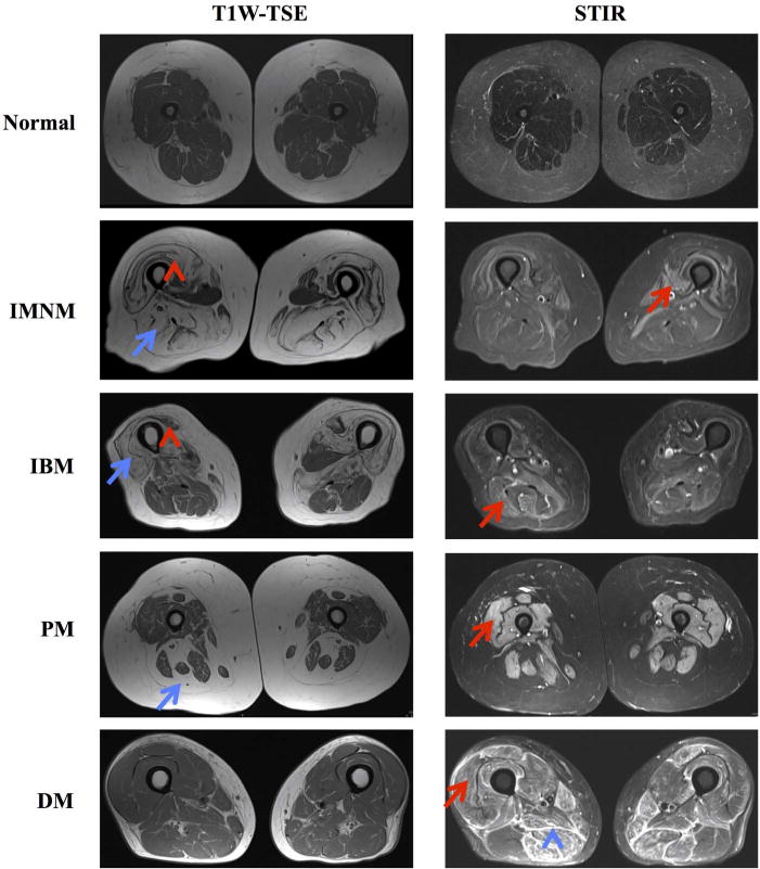
Objectives: The aims of this study were to define the pattern of muscle involvement in patients with immune-mediated necrotising myopathy (IMNM) relative to those with other inflammatory myopathies and to compare patients with IMNM with different autoantibodies.
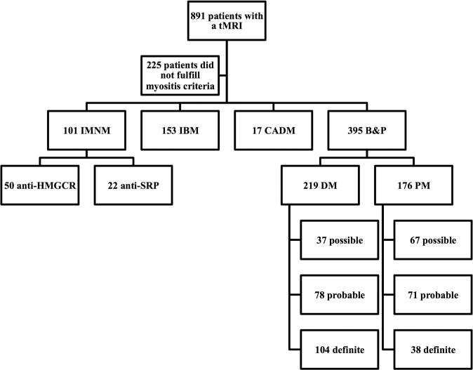
Methods: All Johns Hopkins Myositis Longitudinal Cohort subjects with a thigh MRI (tMRI) who fulfilled criteria for IMNM, dermatomyositis (DM), polymyositis (PM), inclusion body myositis (IBM) or clinically amyopathic DM (CADM) were included in the study. Muscles were assessed for intramuscular and fascial oedema, atrophy and fatty replacement. Disease subgroups were compared using univariate and multivariate analyses. Patients with IMNM with anti-signal recognition particle (SRP) autoantibodies were compared with those with IMNM with anti-HMG-CoA reductase (HMGCR) autoantibodies.
Results: The study included 666 subjects (101 IMNM, 176 PM, 219 DM, 17 CADM and 153 IBM). Compared with DM or PM, IMNM was characterised by a higher proportion of thigh muscles with oedema, atrophy and fatty replacement (p<0.01). Patients with IMNM with anti-SRP had more atrophy (19%, p=0.003) and fatty replacement (18%, p=0.04) than those with anti-HMGCR. In IMNM, muscle abnormalities were especially common in the lateral rotator and gluteal groups. Fascial involvement was most widespread in DM. Fatty replacement of muscle tissue began early during the course of disease in IMNM and the other groups. An optimal combination of tMRI features had only a 55% positive predictive value for diagnosing IMNM.
Conclusions: Compared with patients with DM or PM, IMNM is characterised by more widespread muscle involvement. Anti-SRP-positive patients have more severe muscle involvement than anti-HMGCR-positive patients.
Keywords: Autoantibodies; Dermatomyositis; Magnetic Resonance Imaging; Polymyositis.
Published by the BMJ Publishing Group Limited. For permission to use (where not already granted under a licence) please go to http://www.bmj.com/company/products-services/rights-and-licensing/.
Conflict of interest statement
Figures
References
-
- Dalakas MC. Inflammatory muscle diseases. N Engl J Med. 2015;373:393–4. - PubMed
-
- Del Grande F, Carrino JA, Del Grande M, et al. Magnetic resonance imaging of inflammatory myopathies. Top Magn Reson Imaging. 2011;22:39–43. - PubMed
-
- Garcia J. MRI in inflammatory myopathies. Skeletal Radiol. 2000;29:425–38. - PubMed
-
- Yoshida K, Kurosaka D, Joh K, et al. Fasciitis as a common lesion of dermatomyositis, demonstrated early after disease onset by en bloc biopsy combined with magnetic resonance imaging. Arthritis Rheum. 2010;62:3751–9. - PubMed
-
- Cox FM, Reijnierse M, Van Rijswijk CS, et al. Magnetic resonance imaging of skeletal muscles in sporadic inclusion body myositis. Rheumatology (Oxf) 2011;50:1153–61. - PubMed
Publication types
MeSH terms
Substances
Supplementary concepts
Grants and funding
LinkOut - more resources
Full Text Sources
Other Literature Sources
Medical
