. 2016 Oct 4;113(40):E5886-E5895.
doi: 10.1073/pnas.1604435113. Epub 2016 Sep 20.

Autonomous translational pausing is required for XBP1u mRNA recruitment to the ER via the SRP pathway

Affiliations

Autonomous translational pausing is required for XBP1u mRNA recruitment to the ER via the SRP pathway

Satoshi Kanda et al. Proc Natl Acad Sci U S A. .

Abstract

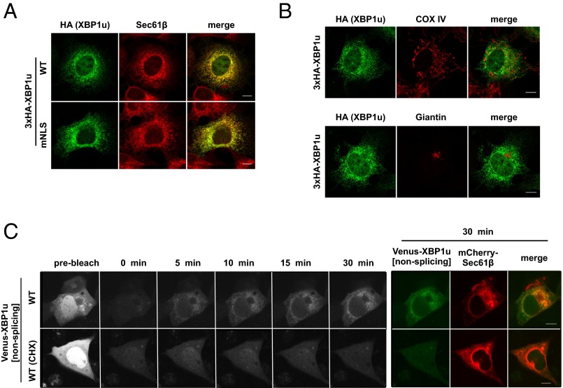
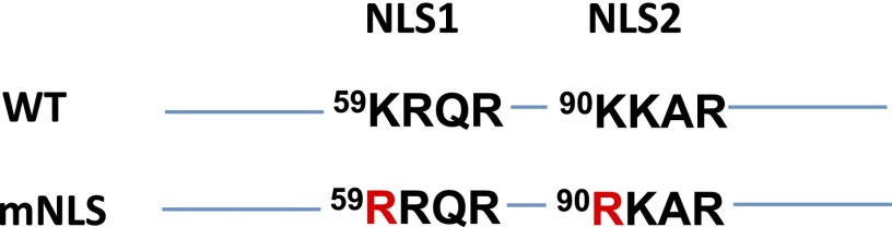
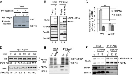
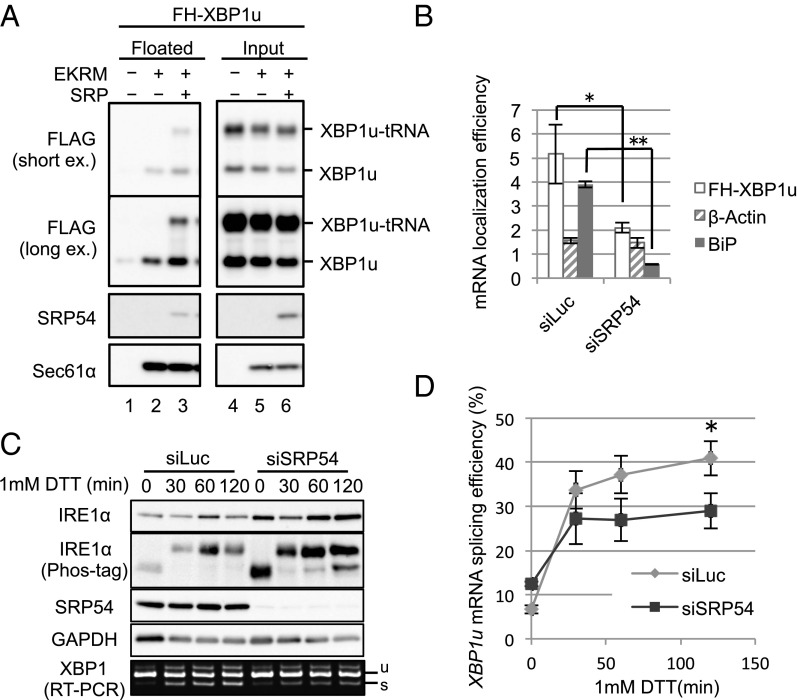
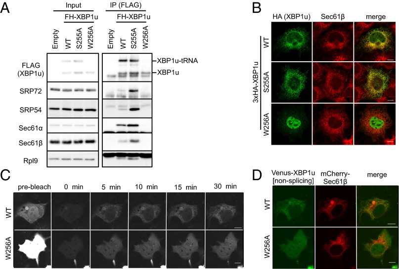
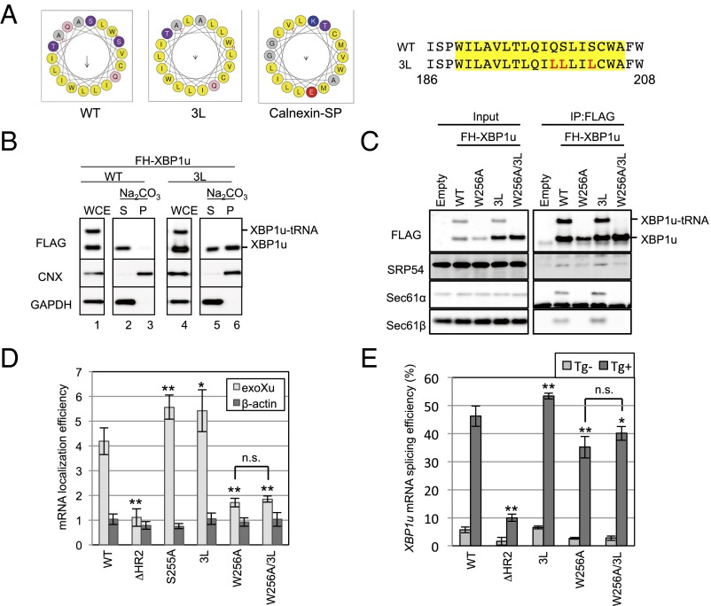
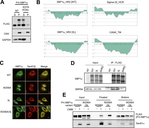
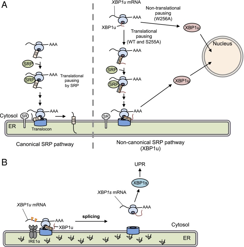
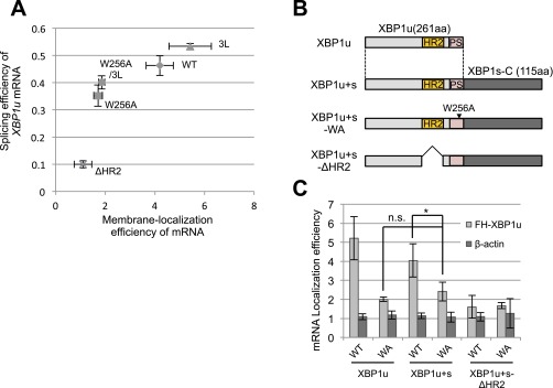
Unconventional mRNA splicing on the endoplasmic reticulum (ER) membrane is the sole conserved mechanism in eukaryotes to transmit information regarding misfolded protein accumulation to the nucleus to activate the stress response. In metazoans, the unspliced form of X-box-binding protein 1 (XBP1u) mRNA is recruited to membranes as a ribosome nascent chain (RNC) complex for efficient splicing. We previously reported that both hydrophobic (HR2) and translational pausing regions of XBP1u are important for the recruitment of its own mRNA to membranes. However, its precise location and the molecular mechanism of translocation are unclear. We show that XBP1u-RNC is specifically recruited to the ER membrane in an HR2- and translational pausing-dependent manner by immunostaining, fluorescent recovery after photobleaching, and biochemical analyses. Notably, translational pausing during XBP1u synthesis is indispensable for the recognition of HR2 by the signal recognition particle (SRP), resulting in efficient ER-specific targeting of the complex, similar to secretory protein targeting to the ER. On the ER, the XBP1u nascent chain is transferred from the SRP to the translocon; however, it cannot pass through the translocon or insert into the membrane. Therefore, our results support a noncanonical mechanism by which mRNA substrates are recruited to the ER for unconventional splicing.

Keywords: SRP; XBP1 mRNA; translational pausing; translocon; unfolded protein response.


Conflict of interest statement

The authors declare no conflict of interest.

Figures

Fig. 1.
Fig. 1.
ER is the initial targeting site of XBP1u. (A and B) HA-XBP1u[nonsplicing] or nuclear localization signal (NLS)-defective mutant (mNLS) HA-XBP1u[mNLS/nonsplicing] transiently expressed in Cos-7 cells was costained with an ER marker (Sec61β), mitochondria marker (COX IV), and Golgi marker (Giantin). (C) Venus-XBP1u[nonsplicing] and mCherry-Sec61β were transiently expressed in Cos-7 cells. The preexisting fluorescence of Venus-XBP1u[nonsplicing] was photobleached, and the emerging fluorescence was observed at the indicated time points. The emerging signal was abrogated by cycloheximide (CHX; 200 μM) treatment for 1 h before the FRAP experiment. The rightmost colored panel shows a merged image of Venus-XBP1u[nonsplicing] and mCherry-Sec61β (ER marker) at 30 min after photobleaching. (Scale bars, 10 μm.)
Fig. S1.
Fig. S1.
Schematic representation of amino acid substitutions in human XBP1u to disrupt two NLS sequences.
Fig. 2.
Fig. 2.
ER targeting machinery for secretory proteins interacts with XBP1u. (A) FLAG-XBP1u-HA was synthesized with RRL in the presence or absence of nontreated or proteinase K (PK)-treated CMMs. Then, CMM-associated proteins were separated from free proteins by ultracentrifugation. P, pellet; S, supernatant. Details regarding the PK treatment of microsomes are provided in Fig. S2. FLAG-XBP1u-HA was detected using an anti-FLAG antibody. (B) Coimmunoprecipitation of SRP54 or translocon components with FLAG-His-XBP1u[nonsplicing] (FH-XBP1u) transiently expressed in HEK293T cells. XBP1u and XBP1u-tRNA indicate full-length and translationally paused FH-XBP1u, respectively. IP, immunoprecipitation. (C) Membrane localization efficiencies of both XBP1u and HR2-deleted XBP1u (ΔHR2) mRNA transiently expressed in HEK293T cells. β-Actin was used as a control for cytosolic mRNA. Bars indicate SD. **P < 0.01 (n = 3) using Student’s t test. (D) Splicing of XBP1u or ΔHR2 mRNA transiently expressed in HEK293T cells was determined by RT-PCR after treatment with thapsigargin (Tg; 0.2 μg/mL) for the indicated times. s, spliced form of XBP1 mRNA; u, unspliced form of XBP1 mRNA. (E) FH-XBP1u[WT] or FH-XBP1u[ΔHR2] translated with RRL was coimmunoprecipitated with SRP54 and ribosomal protein L9 (RPL9) using anti-FLAG antibodies. XBP1u was detected by autoradiography, whereas SRP54 and RPL9 were detected by Western blotting. (F) Coimmunoprecipitation of Sec61β with FH-XBP1u[nonsplicing] transiently expressed in HEK293T cells, which were treated with siRNAs against SRP54 or luciferase (control) for 72 h until the immunoprecipitation assay.
Fig. S2.
Fig. S2.
Supporting information for Fig. 2. (A) Preparation of proteinase K (PK)-treated microsomes. CMMs were treated with the indicated concentration of PK. The efficiency of surface protein digestion was evaluated by the removal of a short cytosolic tail of calnexin. (Right) Membrane topology of calnexin is shown. The full-length calnexin and protected fragment of calnexin were detected using the antibody against the ER-luminal portion of calnexin. (B) XBP1u translation was not affected by PK treatment in the presence of a PK inhibitor. XBP1u was synthesized via the in vitro translation system with mock-treated CMMs or PK-treated CMMs with PMSF (2 mM) and benzamidine (2 mM). (C) Endogenous Sec61β and/or HA-tagged Sec61β was coimmunoprecipitated with FH-XBP1u and FH-XBP1u-tRNA using anti-FLAG antibodies. In contrast, FH-XBP1u and/or FH-XBP1u-tRNA was coimmunoprecipitated with Sec61β and/or Sec61β-HA from the cell lysate derived from HEK293T cells transiently expressing FH-XBP1u and/or Sec61β-HA. Anti-FLAG or anti-HA antibody was used for immunoprecipitation (IP). (D) Coimmunoprecipitation of endogenous Sec61β with FH-XBP1u from the cell lysate derived from HEK293T cells transiently expressing FH-XBP1u. Anti-Sec61β antibody was used for immunoprecipitation.
Fig. 3.
Fig. 3.
SRP recruits XBP1u-RNC to the ER. (A) FH-XBP1u was translated in WGE in the presence or absence of 5 nM purified SRP and CMMs treated with EDTA and high-salt medium (EKRM) to remove preexisting SRP on CMMs. The membrane-bound proteins were separated by a membrane-flotation assay. Proteins in those fractions were detected by immunoblotting. (Top and Middle Top) Same result of immunoblotting exposed for a short time and a long time, respectively, is shown. (B) Membrane localization efficiency (membrane-bound/cytosol) of XBP1u, β-actin, and BiP mRNA in HeLa cells stably expressing FH-XBP1u with SRP54 knockdown for 96 h was quantified as described in Fig. 2C. Bars indicate SD. *P < 0.05, **P < 0.01 (n = 3) in siLuc (Control) vs. siSRP54 using Student’s t test. (C) HeLa cells stably expressing XBP1u-ps with SRP54 knockdown were treated with 1 mM DTT for the indicated times. Western blot analyses of the phosphorylated state of IRE1α and the abundance of indicated proteins are shown. The splicing of XBP1u-ps mRNA was analyzed by RT-PCR. (D) Proportion of the spliced form with respect to the total XBP1u-ps mRNA was calculated from C. Bars indicate SD. *P < 0.05 (n = 3) in siLuc (Control) vs. siSRP54 at 120 min using Student’s t test.
Fig. 4.
Fig. 4.
Translational pausing enables XBP1u-RNC to be a client for the SRP-mediated ER-targeting pathway. (A) Coimmunoprecipitation of FH-XBP1u[WT], FH-XBP1u[S255A], and FH-XBP1u[W256A] with the indicated proteins in the cell lysate derived from HEK293T cells transiently expressing FH-XBP1u[WT] and its variants. FH-XBP1u[S255A] and FH-XBP1u[W256A] are the prolonged- and pausing-defective mutants, respectively. (B) Wild-type and variants of HA-XBP1u[nonsplicing] transiently expressed in Cos-7 cells were costained with endogenous Sec61β. (C and D) FRAP analysis of Venus-XBP1u[WT/nonsplicing] or Venus-XBP1u[W256A/nonsplicing] transiently expressed in Cos-7 cells is shown. (D) Merged image of Venus-XBP1u[WT/nonsplicing] and mCherry-Sec61β (ER marker) at 30 min after photobleaching. The detailed analysis is the same as described in Fig. 1C. (Scale bars, 10 μm.)
Fig. 5.
Fig. 5.
Unusual mode of ER targeting of XBP1u-RNC via SRP. (A) Helical wheel plots of amino acids in HR2 of XBP1u and the calnexin signal peptide are generated by using HeliQuest (heliquest.ipmc.cnrs.fr) (40). Wild-type and 3L mutant XBP1u are indicated as WT and 3L, respectively. (Right) Amino acid sequences of HR2 of XBP1u are shown. Sequences in the yellow rectangle are used for the helical wheel plot. Red characters in the amino acid sequences are substituted amino acids in the 3L mutant. (B) Microsomes derived from HEK293T cells transiently expressing FH-XBP1u and its variants were treated with sodium carbonate to examine the membrane-binding mode of FH-XBP1u variants. Calnexin (CNX) and GAPDH were used as controls for membrane and cytosolic proteins, respectively. (C) Coimmunoprecipitation of FH-XBP1u and its variants with indicated proteins in the cell lysate derived from HEK293T cells transiently expressing FH-XBP1u and its variants. (D) Membrane localization efficiencies of FH-XBP1u mRNA expressed in HEK293T cells were quantified as described in Fig. 2C. Bars indicate SD. *P < 0.05, **P < 0.01 using ANOVA. (E) Splicing efficiencies (percentage of XBP1s mRNA to total XBP1 mRNA) of XBP1u-ps and its variants transiently expressed in HEK293T cells. ER stress was induced with 0.2 μg/mL Tg for 1 h. Bars indicate SD. *P < 0.05, **P < 0.01 using ANOVA. n.s., nonsignificant difference.
Fig. S3.
Fig. S3.
Supporting information for Fig. 5. (A) EDTA and high-salt treatment of microsomes in HEK293T cells transiently expressing wild-type or 3L mutant (3L) FH-XBP1u were analyzed by Western blotting. Calnexin (CNX) and GAPDH were used as the control membrane and cytosolic protein, respectively. (B) Kyte and Doolittle hydrophobicity plots for HR2 of WT and 3L mutant XBP1u (window size: 9) are visualized using a ProtScale analysis implemented in ExPASy (web.expasy.org/protscale/). (C) Variants of FH-XBP1u transiently expressed in HeLa cells were costained with Sec61β. (Scale bars, 10 μm.) (D) FH-XBP1u variants translated with RRL were coimmunoprecipitated with SRP54. XBP1u was detected by autoradiography, whereas SRP54 was detected by immunoblotting. (E) Posttranslational ER-targeting assay of FH-XBP1u[W256A] and FH-XBP1u[W256A/3L]. Total protein before fractionation is shown as the input. Floated and bottom fractions are shown. The presence and absence of the ER membrane (EKRM) are indicated by “−” and “+”, respectively. Additional information is provided in Materials and Methods.
Fig. 6.
Fig. 6.
Working model of translational pausing in SRP-mediated localization of XBP1 mRNA. (A) Canonical SRP pathway (Left) and the noncanonical SRP pathway reported here (Right) are indicated. In the latter case, newly synthesized XBP1u is paused at the C terminus region of the nascent XBP1u polypeptide. Under such conditions, HR2 is located just outside of the ribosomal tunnel and is recognized by SRP. The SRP-bound RNC complex associated with its own XBP1u mRNA is recruited to the translocon via SR. After pausing, XBP1u is completely translated, but cannot be inserted into the ER owing to rejection of the translocon. Rejected XBP1u carrying NLS is quickly transported into the nucleus. In contrast, the pausing-defective mutant XBP1u[W256A], whose HR2 cannot be recognized by SRP, is translated and transported into the nucleus. SS, signal sequence, including the signal anchor. (B) Under ER stress, XBP1u mRNA associated with paused RNC on the translocon is efficiently spliced by activated IRE1α, leading to production of the active transcription factor XBP1s, which up-regulates unfolded protein response (UPR) target genes to mitigate ER stress.
Fig. S4.
Fig. S4.
Splicing efficiency and membrane localization efficiency of XBP1u mRNA. (A) Relationship between splicing efficiency and membrane localization efficiency of XBP1u mRNA according to Figs. 2C and 5 D and E is shown. The bar indicates SD. (B) Primary structure of variants of XBP1u and C-terminal extended XBP1u, XBP1u+s. PS, pausing sequence; WA, W256A. (C) Membrane localization efficiencies of XBP1u variant mRNA transiently expressed in HEK293T cells were quantified as described in Fig. 2C. Bars indicate SD. *P < 0.05 (n = 4) using ANOVA. n.s., nonsignificant difference.

Similar articles

Cited by

References

    1. Walter P, Blobel G. Translocation of proteins across the endoplasmic reticulum III. Signal recognition protein (SRP) causes signal sequence-dependent and site-specific arrest of chain elongation that is released by microsomal membranes. J Cell Biol. 1981;91(2 Pt 1):557–561. - PMC - PubMed
    1. Akopian D, Shen K, Zhang X, Shan SO. Signal recognition particle: An essential protein-targeting machine. Annu Rev Biochem. 2013;82:693–721. - PMC - PubMed
    1. Keenan RJ, Freymann DM, Stroud RM, Walter P. The signal recognition particle. Annu Rev Biochem. 2001;70:755–775. - PubMed
    1. Lakkaraju AK, Mary C, Scherrer A, Johnson AE, Strub K. SRP keeps polypeptides translocation-competent by slowing translation to match limiting ER-targeting sites. Cell. 2008;133(3):440–451. - PMC - PubMed
    1. Halic M, et al. Structure of the signal recognition particle interacting with the elongation-arrested ribosome. Nature. 2004;427(6977):808–814. - PubMed

Publication types

MeSH terms