. 2016 Nov;22(11):1739-1749.
doi: 10.1261/rna.058461.116. Epub 2016 Sep 20.

Mutually exclusive RNA secondary structures regulate translation initiation of DinQ in Escherichia coli

Affiliations

Mutually exclusive RNA secondary structures regulate translation initiation of DinQ in Escherichia coli

Knut I Kristiansen et al. RNA. 2016 Nov.

Abstract

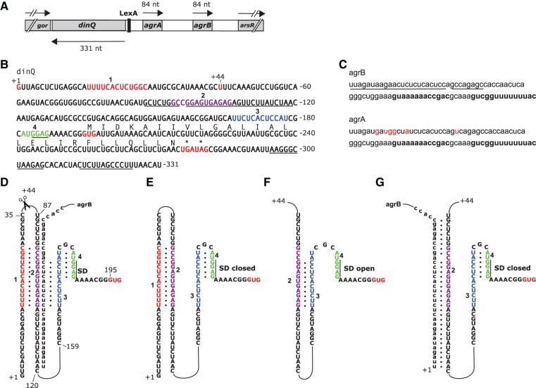
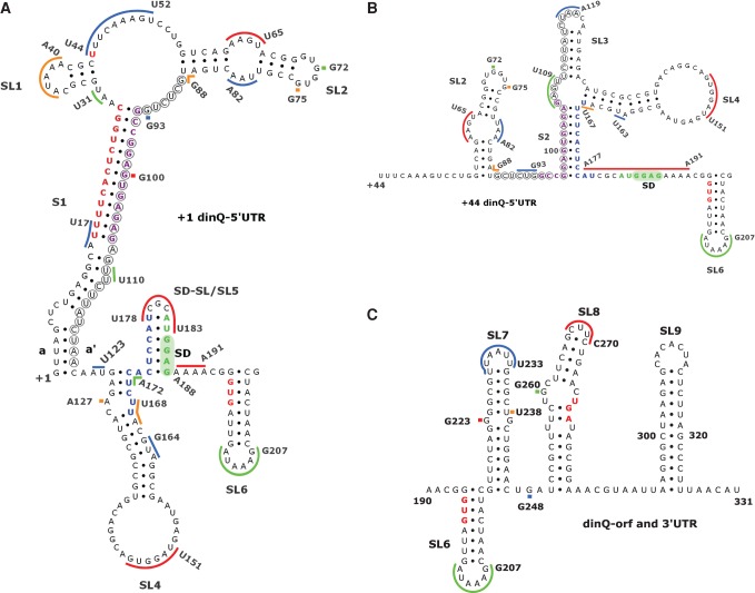
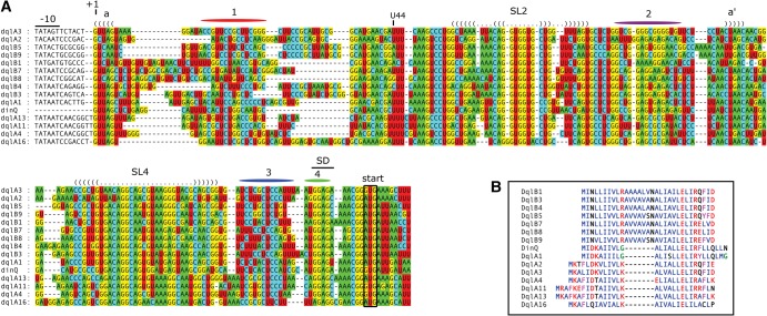
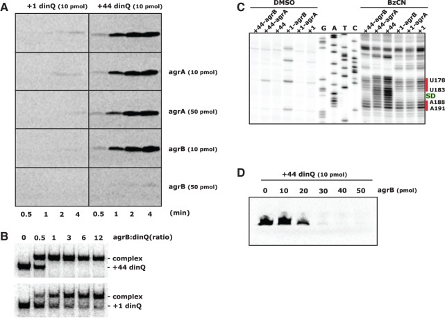
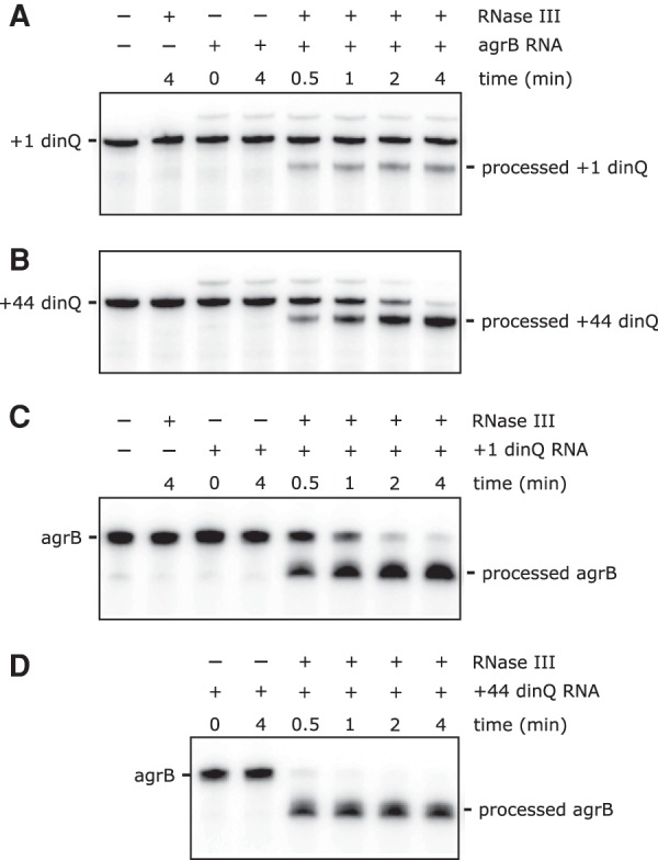
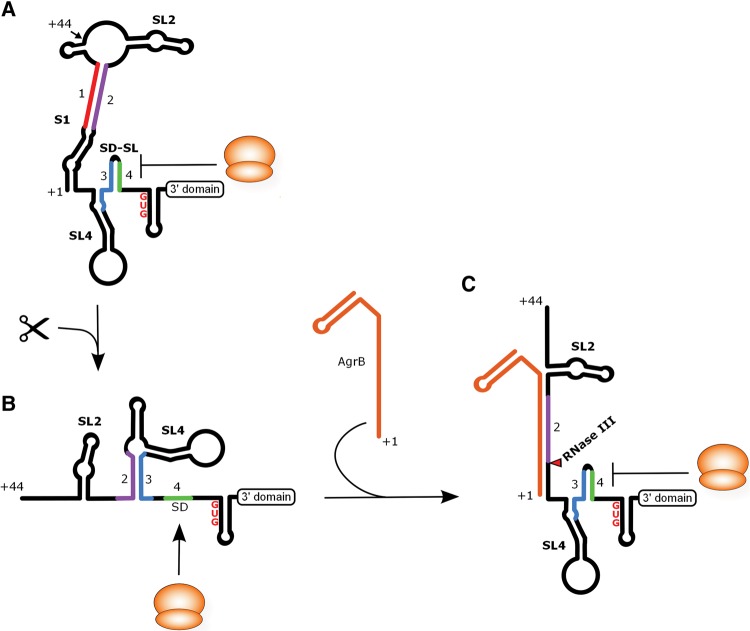
Protein translation can be affected by changes in the secondary structure of mRNA. The dinQ gene in Escherichia coli encodes a primary transcript (+1) that is inert to translation. Ribonucleolytic removal of the 44 first nucleotides converts the +1 transcript into a translationally active form, but the mechanism behind this structural change is unknown. Here we present experimental evidence for a mechanism where alternative RNA secondary structures in the two dinQ mRNA variants affect translation initiation by mediating opening or closing of the ribosome binding sequence. This structural switch is determined by alternative interactions of four sequence elements within the dinQ mRNA and also by the agrB antisense RNA. Additionally, the structural conformation of +1 dinQ suggests a locking mechanism comprised of an RNA stem that both stabilizes and prevents translation initiation from the full-length dinQ transcript. BLAST search and multiple sequence alignments define a new family of dinQ-like genes widespread in Enterobacteriaceae with close RNA sequence similarities in their 5' untranslated regions. Thus, it appears that a whole new family of genes is regulated by the same mechanism of alternative secondary RNA structures.

Keywords: DinQ; E. coli; RNA processing; RNA structure; translation initiation.


Figures

FIGURE 1.
FIGURE 1.
(A) A schematic representation of the dinQ, agrA, and agrB genes and transcripts. Drawing is to scale. The positions of the LexA binding site and the flanking gor and arsR genes are shown. (B) dinQ mRNA. Transcriptional initiation site +1 and processing site +44 are indicated (red letters). RNA sequences 1, 2, 3, and 4 are indicated by red, purple, blue, and green letters, respectively. Underlined are the agrB antisense region, the Shine-Dalgarno (SD) sequence, and the dinQ terminator. DinQ start and stop codons (asterisks) are shown in red. DinQ translation sequence is shown above the nucleotide sequence. All numbering of the dinQ sequence and RNA structures are relative to the +1 transcription initiation site. (C) agrB and agrA RNA transcripts. Antisense sequence is underlined (agrB). Rho-independent transcriptional terminator is indicated in bold. Red letters indicate nucleotides in agrA that are different from agrB. (D) Potential base-pairing of four RNA sequences and agrB antisense RNA: (red) sequence 1 (U16–C28); (purple) sequence 2 (G94–G106); (blue) sequence 3 (U167–U178); (green) sequence 4 (A182–G187). The 36 first nucleotides of agrB antisense RNA are shown base-paired with G88–A119 encompassing sequence 2 of dinQ. The +44 processing site, the Shine-Dalgarno (SD) sequence (G184–G187), and the GUG start codon starting at G195 are indicated. (E) +1 dinQ mRNA. Base-pairing of sequences 1 and 2 allow sequences 3 and 4 to form the closed SD hairpin. (F) +44 dinQ mRNA. Elimination of sequence 1 allows formation of the competing duplex 2:3, thus excluding sequences 3 and 4 to form the closed SD hairpin. (G) Binding of agrB to G88–A119 in +44 dinQ sequestrates sequence 2, allowing the closed SD hairpin to form.
FIGURE 2.
FIGURE 2.
Secondary structure models of dinQ RNA. (A) +1 dinQ 5′ UTR. (B) Secondary structure of +44 dinQ 5′ UTR. (C) Secondary structure of the dinQ-ORF and 3′ UTR region in +1 dinQ and +44 dinQ RNA. Different colored bars correspond to SHAPE reactive regions and match Supplemental Figure 1. Nucleotides in red, purple, blue, and green correspond to nucleotide sequences 1, 2, 3, and 4, respectively. Sequence interaction a/a′ supported by sequence conservation is indicated. Nucleotides encircled indicate the agrB antisense region in dinQ. Stem–loops (SL) and stems (S) are numbered, the Shine-Dalgarno (SD) region is shaded green, and the GUG translational start, UGA stop, and U44 cleavage site are shown in red.
FIGURE 3.
FIGURE 3.
Conserved sequence and structure elements in the 5′ UTR of dinQ and dql genes. (A) Nucleotide positions are relative to the dinQ mRNA sequence. Conserved −10 promoter elements, Shine-Dalgarno (SD) sequences, and start codons are indicated. Nucleotide regions colored red, purple, blue, and green correspond to nucleotide sequences 1, 2, 3, and 4, respectively. Conservation of stem–loops 2 and 4 (SL2 and SL4) is indicated with a dot-bracket notation. Conserved upper- and lower stems are indicated with a dot-bracket representation and colored nucleotides according to standard IUPAC nucleotide annotation. Sequence interaction a/a′ supported by sequence conservation is indicated. (B) Sequence alignment of DinQ and 14 Dql peptides. Hydrophobic amino acids (AILMV) are blue; polar amino acids (DEKR) are red; aromatic amino acids (FY) are magenta; small amino acids (G) are green; all others (CNPQST) are black.
FIGURE 4.
FIGURE 4.
Translational capacity of dinQ transcripts in the presence of agrB RNA. (A) In vitro translation in S30 extracts of +1 dinQ and +44 dinQ RNA in the absence or presence of agrA or agrB antisense RNA. Amount of RNA and incubation time is indicated. (B) Complex formation between labeled +44 dinQ and unlabeled agrB RNA (upper autoradiograph), and labeled +1 dinQ and unlabeled agrB RNA (lower autoradiograph). The electrophoretic mobility shift assay was performed as described in Materials and Methods using 1.3 pmol of labeled +1 dinQ or +44 dinQ RNA mixed with different concentrations of agrB RNA in a final volume of 15 µL. The ratios of agrB and dinQ RNA are indicated. (C) SHAPE reactivity of the Shine-Dalgarno (SD) region of +1 dinQ and +44 dinQ RNA monitored with or without a fivefold excess of agrB or agrA RNA. The SD sequence G184–G187 is indicated in green. Two red bars correspond to SHAPE reactive regions of the SD region in +1 dinQ and +44 dinQ. (D) In vitro translation in S30 extracts of +44 dinQ RNA mixed with increasing amounts of agrB antisense RNA. Incubation time is 4 min.
FIGURE 5.
FIGURE 5.
RNase III-mediated cleavage of dinQ and agrB RNA. (A,B) 4.4 pmol of 3′ end labeled +1 dinQ or +44 dinQ RNA, alone or mixed with a twofold excess of antisense agrB RNA in a final volume of 17.5 µL, was treated with 0.001 U of RNase III for different times as indicated. (C,D) 6.6 pmol of 3′ end labeled agrB RNA, alone or mixed with an equimolar amount of +1 dinQ or +44 dinQ RNA in a final volume of 26.3 µL, was treated with 0.005 U of RNase III for different times as indicated.
FIGURE 6.
FIGURE 6.
A model of dinQ translation regulation based on the data presented. (A) Secondary structure of the 5′ UTR of +1 dinQ. The identical 3′ domain structure of +1 dinQ and +44 dinQ RNA is indicated with a labeled box. The ribosome is drawn as an orange sphere. The SL2, SL4, SD, and SD-SL are labeled. SL = Stem–loop, SD = Shine-Dalgarno. (B) Secondary structure of the 5′ UTR of +44 dinQ RNA. (C) The translationally inactive state of +44 dinQ RNA when hybridized to agrB RNA. The site of RNase III cleavage of dinQ is indicated with a red triangle.

References

    1. Alix E, Blanc-Potard AB. 2009. Hydrophobic peptides: novel regulators within bacterial membrane. Mol Microbiol 72: 5–11. - PubMed
    1. Brantl S, Jahn N. 2015. sRNAs in bacterial type I and type III toxin-antitoxin systems. FEMS Microbiol Rev 39: 413–427. - PubMed
    1. Court DL, Gan J, Liang YH, Shaw GX, Tropea JE, Costantino N, Waugh DS, Ji X. 2013. RNase III: genetics and function; structure and mechanism. Annu Rev Genet 47: 405–431. - PMC - PubMed
    1. Darfeuille F, Unoson C, Vogel J, Wagner EG. 2007. An antisense RNA inhibits translation by competing with standby ribosomes. Mol Cell 26: 381–392. - PubMed
    1. Darty K, Denise A, Ponty Y. 2009. VARNA: interactive drawing and editing of the RNA secondary structure. Bioinformatics 25: 1974–1975. - PMC - PubMed

LinkOut - more resources