Radioprotective effects of hesperidin on oxidative damages and histopathological changes induced by X-irradiation in rats heart tissue
- PMID: 27651565
- PMCID: PMC5019037
- DOI: 10.4103/0971-6203.189482
Radioprotective effects of hesperidin on oxidative damages and histopathological changes induced by X-irradiation in rats heart tissue
Abstract
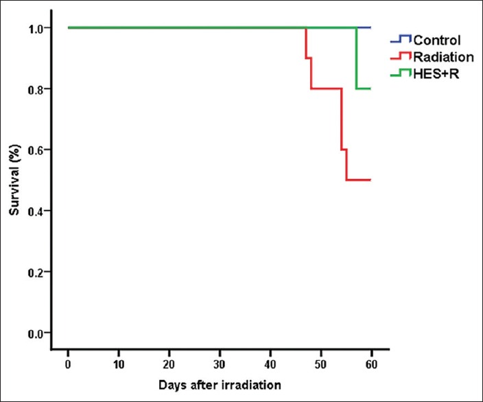
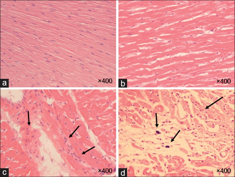
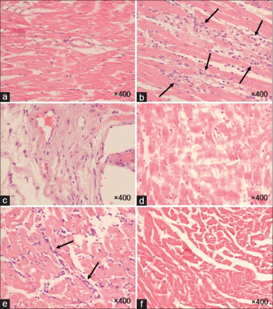
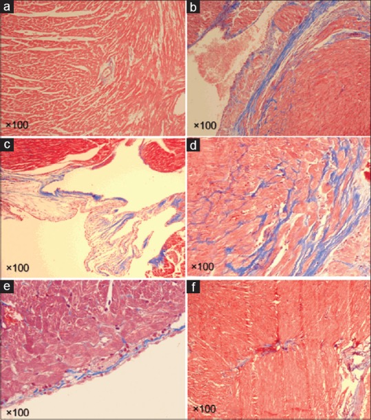
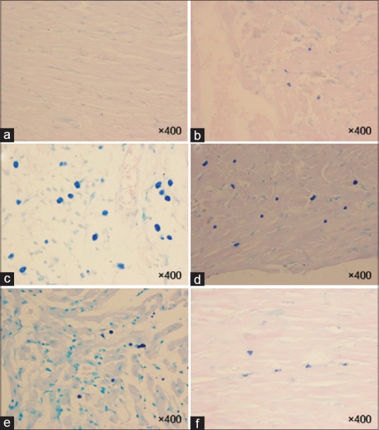
This study was carried out to evaluate radioprotective effects of hesperidin (HES) administration before the irradiation on the cardiac oxidative stress and histopathological changes in an experimental rat model. The cardiovascular complications of radiation exposure cause morbidity and mortality in patients who received radiotherapy. HES, an antioxidant flavonoid found in citrus fruits, suggests the protection against the tissue damage. Fifty-eight rats were divided into four groups: Group 1 received phosphate buffered saline (PBS) and sham radiation; Group 2, HES and sham radiation; Group 3, PBS and radiation; and Group 4, HES and radiation. The rats were exposed to single dose of 18 Gy of 6 MV X-ray. One hundred milligrams per kilogram doses of HES was administered for 7 days before irradiation. The estimation of superoxide dismutase (SOD), malondialdehyde (MDA), and histopathological analyses was performed at 24 h and 8 weeks after radiation exposure. The irradiation of chest area resulted in an elevated MDA level and decreased SOD activity. Moreover, long-term pathological lesions of radiation were inflammation, fibrosis, the increased number of mast cells and macrophages, and development of plaque, vascular leakage, myocardial degeneration, and myocyte necrosis. Although the administration of HES decreases inflammation, fibrosis, mast cell and macrophage numbers, and myocyte necrosis, it did not result in reduced thrombus, myocardium degeneration, and vascular leakage. In conclusion, these results suggest that HES can perform a radioprotection action. The protective effect of HES may be attributable to its immunomodulatory effects and free radical-scavenging properties.
Keywords: Cardiomyopathy; cardiotoxicity; hesperidin; radioprotector.
Figures
References
-
- Adams MJ, Hardenbergh PH, Constine LS, Lipshultz SE. Radiation-associated cardiovascular disease. Crit Rev Oncol Hematol. 2003;45:55–75. - PubMed
-
- Madan R, Benson R, Sharma DN, Julka PK, Rath GK. Radiation induced heart disease: Pathogenesis, management and review literature. J Egypt Natl Canc Inst. 2015;27:187–93. - PubMed
-
- Heidenreich PA, Schnittger I, Strauss HW, Vagelos RH, Lee BK, Mariscal CS, et al. Screening for coronary artery disease after mediastinal irradiation for Hodgkin's disease. J Clin Oncol. 2007;25:43–9. - PubMed
-
- Carver JR, Shapiro CL, Ng A, Jacobs L, Schwartz C, Virgo KS, et al. American Society of Clinical Oncology clinical evidence review on the ongoing care of adult cancer survivors: Cardiac and pulmonary late effects. J Clin Oncol. 2007;25:3991–4008. - PubMed
-
- Shimizu Y, Pierce DA, Preston DL, Mabuchi K. Studies of the mortality of atomic bomb survivors. Report 12, part II. Noncancer mortality: 1950-1990. Radiat Res. 1999;152:374–89. - PubMed
LinkOut - more resources
Full Text Sources
Other Literature Sources