

Protective mechanisms of microRNA-27a against oxygen-glucose deprivation-induced injuries in hippocampal neurons
- PMID: 27651777
- PMCID: PMC5020828
- DOI: 10.4103/1673-5374.189194
Protective mechanisms of microRNA-27a against oxygen-glucose deprivation-induced injuries in hippocampal neurons
Abstract
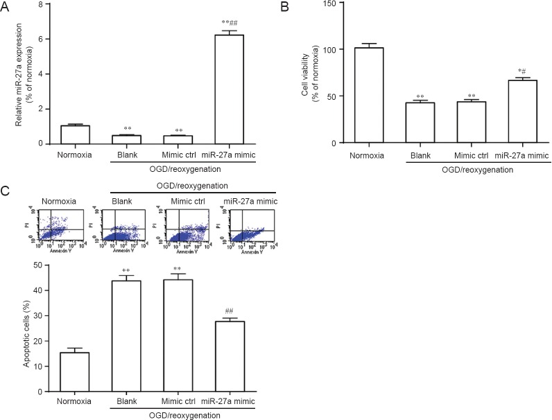
Hypoxic injuries during fetal distress have been shown to cause reduced expression of microRNA-27a (miR-27a), which regulates sensitivity of cortical neurons to apoptosis. We hypothesized that miR-27a overexpression attenuates hypoxia- and ischemia-induced neuronal apoptosis by regulating FOXO1, an important transcription factor for regulating the oxidative stress response. miR-27a mimic was transfected into hippocampal neurons to overexpress miR-27a. Results showed increased hippocampal neuronal viability and decreased caspase-3 expression. The luciferase reporter gene system demonstrated that miR-27a directly binded to FOXO1 3'UTR in hippocampal neurons and inhibited FOXO1 expression, suggesting that FOXO1 was the target gene for miR-27a. These findings confirm that miR-27a protects hippocampal neurons against oxygen-glucose deprivation-induced injuries. The mechanism might be mediated by modulation of FOXO1 and apoptosis-related gene caspase-3 expression.
Keywords: FOXO1; apoptosis; brain injury; caspase 3; cell survival; hippocampal neurons; hypoxic-ischemic; luciferase reporter gene system; miR-27a; nerve regeneration; neural regeneration; neuroprotection; oxygen-glucose deprivation.
Conflict of interest statement
Conflicts of Interest: None declared.
Figures


Similar articles
-
MicroRNA-27a Promotes Oxidative-Induced RPE Cell Death through Targeting FOXO1.Biomed Res Int. 2021 Nov 1;2021:6666506. doi: 10.1155/2021/6666506. eCollection 2021. Biomed Res Int. 2021. PMID: 34761005 Free PMC article.
-
MicroRNA-132 protects hippocampal neurons against oxygen-glucose deprivation-induced apoptosis.Int J Immunopathol Pharmacol. 2017 Sep;30(3):253-263. doi: 10.1177/0394632017715837. Epub 2017 Jun 19. Int J Immunopathol Pharmacol. 2017. Retraction in: Int J Immunopathol Pharmacol. 2021 Jan-Dec;35:20587384211040388. doi: 10.1177/20587384211040388. PMID: 28627974 Free PMC article. Retracted.
-
MicroRNA-27a-3p Downregulation Inhibits Inflammatory Response and Hippocampal Neuronal Cell Apoptosis by Upregulating Mitogen-Activated Protein Kinase 4 (MAP2K4) Expression in Epilepsy: In Vivo and In Vitro Studies.Med Sci Monit. 2019 Nov 11;25:8499-8508. doi: 10.12659/MSM.916458. Med Sci Monit. 2019. PMID: 31710596 Free PMC article.
-
N-hydroxy-N'-(4-butyl-2-methylphenyl)-formamidine attenuates oxygen-glucose deprivation and reoxygenation-induced cerebral ischemia-reperfusion injury via regulation of microRNAs.J Integr Neurosci. 2020 Jun 30;19(2):303-311. doi: 10.31083/j.jin.2020.02.1236. J Integr Neurosci. 2020. PMID: 32706194
-
Neuroprotective effects of miR-27a against traumatic brain injury via suppressing FoxO3a-mediated neuronal autophagy.Biochem Biophys Res Commun. 2017 Jan 22;482(4):1141-1147. doi: 10.1016/j.bbrc.2016.12.001. Epub 2016 Dec 2. Biochem Biophys Res Commun. 2017. PMID: 27919684
Cited by
-
miR-27a protects against acute lung injury in LPS-treated mice by inhibiting NF-κB-mediated inflammatory response.Int J Clin Exp Pathol. 2018 Jun 1;11(6):2980-2989. eCollection 2018. Int J Clin Exp Pathol. 2018. PMID: 31938423 Free PMC article.
-
[Yiqihuoxue prescription promotes nerve regeneration by miR-124-mediated regulation of Wnt signaling in rats].Nan Fang Yi Ke Da Xue Xue Bao. 2017 Aug 20;37(8):1047-1053. doi: 10.3969/j.issn.1673-4254.2017.08.08. Nan Fang Yi Ke Da Xue Xue Bao. 2017. PMID: 28801284 Free PMC article. Chinese.
-
MicroRNA-27a alleviates LPS-induced acute lung injury in mice via inhibiting inflammation and apoptosis through modulating TLR4/MyD88/NF-κB pathway.Cell Cycle. 2018;17(16):2001-2018. doi: 10.1080/15384101.2018.1509635. Epub 2018 Sep 19. Cell Cycle. 2018. PMID: 30231673 Free PMC article.
-
Blood microRNAs as potential diagnostic markers for hemorrhagic stroke.Neural Regen Res. 2017 Jan;12(1):13-18. doi: 10.4103/1673-5374.198965. Neural Regen Res. 2017. PMID: 28250731 Free PMC article. Review.
-
Circular RNA expression profiles in neonatal rats following hypoxic-ischemic brain damage.Int J Mol Med. 2019 Apr;43(4):1699-1708. doi: 10.3892/ijmm.2019.4111. Epub 2019 Feb 26. Int J Mol Med. 2019. PMID: 30816430 Free PMC article.
References
-
- Beppu K, Sasaki T, Tanaka KF, Yamanaka A, Fukazawa Y, Shigemoto R, Matsui K. Optogenetic countering of glial acidosis suppresses glial glutamate release and ischemic brain damage. Neuron. 2014;81:314–320. - PubMed
-
- Berger HR, Morken TS, Vettukattil R, Brubakk AM, Sonnewald U, Wideroe M. No improvement of neuronal metabolism in the reperfusion phase with melatonin treatment after hypoxic-ischemic brain injury in the neonatal rat. J Neurochem. 2016;136:339–350. - PubMed
LinkOut - more resources
Full Text Sources
Other Literature Sources
Research Materials
Miscellaneous
