. 2016 Sep 13;8(1):96.
doi: 10.1186/s13148-016-0263-9. eCollection 2016.

Molecular Pap smear: HPV genotype and DNA methylation of ADCY8, CDH8, and ZNF582 as an integrated biomarker for high-grade cervical cytology

Affiliations

Molecular Pap smear: HPV genotype and DNA methylation of ADCY8, CDH8, and ZNF582 as an integrated biomarker for high-grade cervical cytology

Jane Shen-Gunther et al. Clin Epigenetics. .

Abstract

Background: The Pap smear has remained the foundation for cervical cancer screening for over 70 years. With advancements in molecular diagnostics, primary high-risk human papillomavirus (hrHPV) screening has recently become an accepted stand-alone or co-test with conventional cytology. However, both diagnostic tests have distinct limitations. The aim of this study was to determine the association between HPV genotypes and cellular epigenetic modifications in three grades of cervical cytology for screening biomarker discovery.

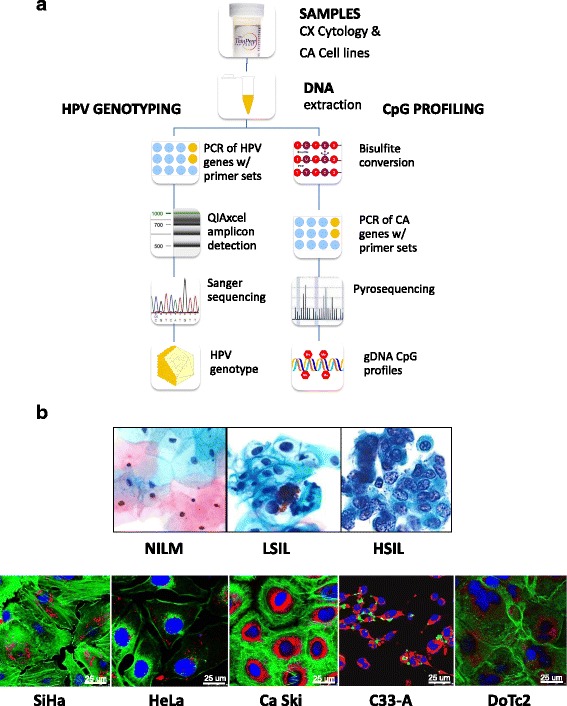
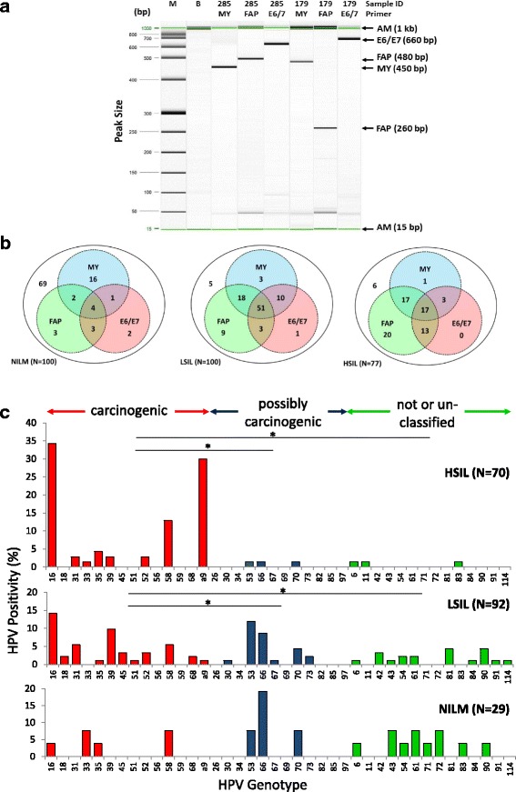
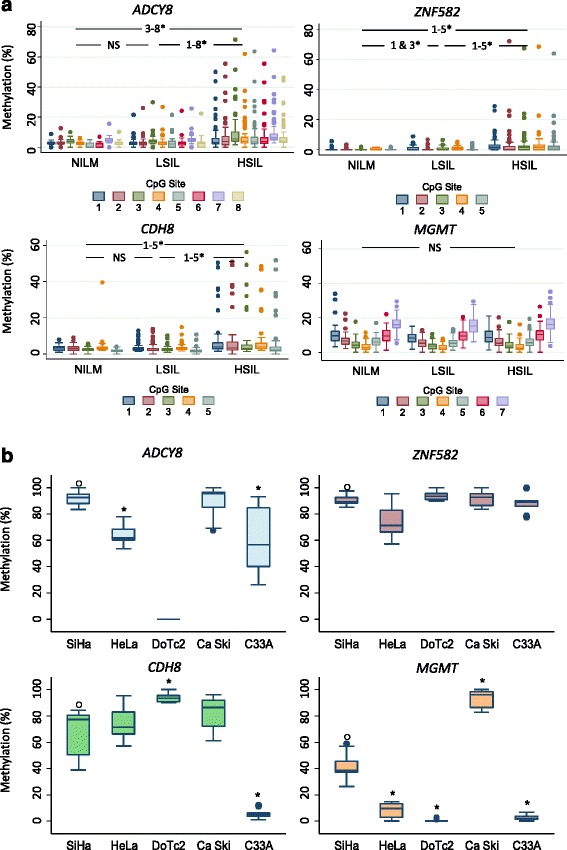
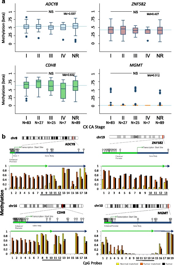
Methods: This prospective, cross-sectional study used residual liquid-based cytology samples for HPV genotyping and epigenetic analysis. Extracted DNA was subjected to parallel polymerase chain reactions using three primer sets (MY09/11, FAP59/64, E6-E7 F/B) for HPV DNA amplification. HPV+ samples were genotyped by DNA sequencing. Promoter methylation of four candidate tumor suppressor genes (adenylate cyclase 8 (ADCY8), cadherin 8, type 2 (CDH8), MGMT, and zinc finger protein 582 (ZNF582)) out of 48 genes screened was quantified by bisulfite-pyrosequencing of genomic DNA. Independent validation of methylation profiles was performed by analyzing data from cervical cancer cell lines and clinical samples from The Cancer Genome Atlas (TCGA).

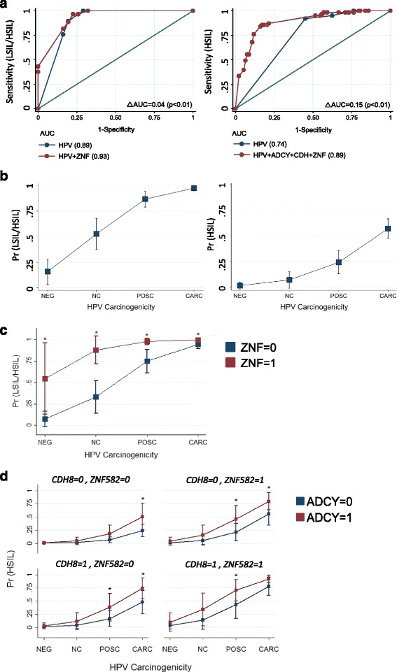
Results: Two hundred seventy-seven quality cytology samples were analyzed. HPV was detected in 31/100 (31 %) negative for intraepithelial lesion or malignancy (NILM), 95/100 (95 %) low-grade squamous intraepithelial lesion (LSIL), and 71/77 (92 %) high-grade squamous intraepithelial lesion (HSIL) samples. The proportion of IARC-defined carcinogenic HPV types in sequenced samples correlated with worsening grade: NILM 7/29 (24 %), LSIL 53/92 (58 %), and HSIL 65/70 (93 %). Promoter methylation of ADCY8, CDH8, and ZNF582 was measured in 170 samples: NILM (N = 33), LSIL (N = 70), and HSIL (N = 67) also correlated with worsening grade. Similar hypermethylation patterns were found in cancer cell lines and TCGA samples. The combination of four biomarkers, i.e., HPV genotype and three-gene promoter methylation, predicted HSIL (AUC 0.89) better than HPV alone (AUC 0.74) by logistic regression and probabilistic modeling.

Conclusions: HPV genotype and DNA methylation of ADCY8, CDH8, and ZNF582 are correlated with cytological grade. Collectively, these biomarkers may serve as a molecular classifier of Pap smears.

Keywords: DNA methylation; HPV; HPV genotyping; Molecular biomarkers; Molecular diagnostics; Pap smear; Pyrosequencing.


Figures

Fig. 1
Fig. 1
Protocol schema and representative images of cervical cytology and cervical carcinoma cell lines used in the study. a Sample collection, DNA extraction, HPV genotyping by Sanger sequencing, and CpG profiling of gene-specific promoters by pyrosequencing. b Three categories of liquid-based cervical cytology: negative for intraepithelial lesion or malignancy (NILM), low-grade squamous intraepithelial lesion (LSIL), and high-grade intraepithelial lesion (HSIL), reveal progressive nuclear enlargement, nuclear membrane irregularity, and chromatin coarseness associated with worsening grade. Five cervical carcinoma cell lines: SiHa, HeLa, Ca Ski, C33-A, and DoTc2, with distinct cytomorphologic features, e.g., cell size and shape, nucleus (blue), nuclear-to-cytoplasmic ratio, chromatin patterns, actin cytoskeleton (green), and mitochondria (red). Each cell line was immunofluorescence labeled and imaged by confocal microscopy (×63 objective). Abbreviations: CX cervical, CA cancer, PCR polymerase chain reaction, HSIL high-grade squamous intraepithelial lesion, LSIL low-grade squamous intraepithelial lesion, NILM negative for intraepithelial lesion or malignancy
Fig. 2
Fig. 2
PCR amplification of HPV DNA by three consensus primer sets and HPV genotyping by amplicon sequencing. a Representative gel image of PCR amplicon detection by high-resolution capillary gel electrophoresis. Representative samples #285 (LSIL) and #179 (HSIL) reveal MY09/11, FAP59/64, and GP-E6/E7 F/B amplicons with expected yield of ~450-, 480- (or 260-bp fragment), and 660-bp fragments, respectively. b Parallel PCR testing for HPV by three primer sets. Venn diagrams show intersecting and complementary sets of cytological samples (N) detected of HPV DNA by MY-, FAP-, and E6/E7 primer sets according to cytological diagnoses, i.e., NILM, LSIL, and HSIL. The net positivity of simultaneous testing for HPV (union of the circles) in NILM, LSIL, and HSIL are 31/100 (31 %), 95/100 (95 %), and 71/77 (92 %), respectively. c HPV genotype distribution of 191 cytology samples with PCR-detected HPV DNA according to cytological diagnoses: NILM, LSIL, and HSIL. The increase in carcinogenic HPV genotypes was coincident with cytological grade (Spearman’s ρ = 0.658, p < 0.001). Samples positive for the 260-bp fragment that aligned closest to HPV-58 were assigned as “alpha-9” species because of the non-specific short sequence length. *p < 0.05 by the chi-square test. Abbreviations: AM alignment marker, B buffer, bp base pair, HSIL high-grade squamous intraepithelial lesion, IARC International Agency for Research on Cancer, LSIL low-grade squamous intraepithelial lesion, M molecular weight ladder, NILM negative for intraepithelial lesion or malignancy
Fig. 3
Fig. 3
Promoter methylation differences in cervical cytology and cervical carcinoma cell lines. a Methylation (%) of total genomic DNA in three grades of cervical cytology, i.e., NILM (N = 33), LSIL (N = 70), and HSIL (N = 67), was compared by CpG positions among four genes (ADCY8, CDH8, ZNF582, and MGMT). Pairwise comparisons of methylation for each CpG position between cytological grades (NILM vs. LSIL, LSIL vs. HSIL, and NILM vs. HSIL) revealed significantly higher levels for HSIL vs. LSIL and LSIL vs. NILM at multiple positions for ADCY8, CDH8, and ZNF582. For MGMT, methylation levels were not significantly different among cytological grades. Methylation levels for each CpG position increased concurrently with cytological grade for ADCY8, CDH8, and ZNF582 by Spearman’s ρ (p < 0.001). *p < 0.05 by the Wilcoxon rank-sum test. b SiHa, HeLa, and Ca Ski cell lines with genome-integrated HPV demonstrated promoter hypermethylation of ADCY8, CDH8, and ZNF582 genes. For HPV-negative cell lines, DoTc2 and C33-A revealed an inconsistent pattern of hypermethylation in the studied genes. Using SiHa methylation (%) as a reference (o), cell lines with significantly different levels are indicated by an asterisk. *p < 0.05 by the Wilcoxon rank-sum test. NS not statistically significant. Cell lines were analyzed for CpG methylation in duplicate collections
Fig. 4
Fig. 4
Promoter methylation of ADCY8, CDH8, ZNF582, and MGMT in the TCGA cervical cancer cohort. a Box plots of CpG methylation (β value) according to FIGO stage for 231 patient samples with squamous cell carcinoma. Gene-specific median methylation values for all FIGO stages are specified (Md) and indicated by the blue reference lines. NS not statistically significant, Kruskal-Wallis P > 0.05. NR stage not reported. b Differential CpG methylation (β value) ante- and post-transcription start site for 257 cervical carcinomas (squamous, N = 231; adenocarcinoma, N = 26) and 3 tumor/matched normal samples. The four panels display the chromosomal positions of ADCY8, CDH8, ZNF582, and MGMT (red line) with an expanded area showing the CpG probes on the Illumina HumanMethylation 450 K microarray (gene ball-and-stick diagrams). The bar graphs present the median DNA methylation (β value) of 257 tumors (black) and 3 matched tumor (orange)/normal (yellow) samples across the ordered CpG probes. The promoter methylation levels were notably higher (~×10) for tumor (median β ~0.6) than the normal samples (median β ~0.06) for ADCY8, CDH8, and ZNF582. The enhancer/promoter and gene body regions are indicated by the green and blue arrows, respectively. The CpG regions selected for bisulfite pyrosequencing of cytology samples are denoted by the underscored CpG probes. The chromosome coordinates for the CpG probes along the X-axis are as follows: ADCY8 (chr8: 132,053,823-131,896,788), CDH8 (chr16: 62,070,072-61,871,849), ZNF582 (chr19: 56,905,383-56,901,457), and MGMT (chr10: 131,264,840-131,304,833). [Chromosome ideograms adapted from NCBI Map Viewer (www.ncbi.nlm.nih.gov/genome/guide/human)]
Fig. 5
Fig. 5
Regression models and predicted probability plots for cytological grades. a Receiver operating characteristic curve analysis using cut-points derived from univariate ROC analysis of gene-specific methylation levels. Multivariable modeling revealed the best predictor to differentiate between NILM and LSIL/HSIL was HPV carcinogenicity and ZNF582_7th CpG position binarized as follows: <1.1 (0), ≥1.1 (1) (ROC AUC = 0.93). For differentiating between NILM/LSIL and HSIL cytology, the best multivariate predictor was the combination of HPV carcinogenicity, ADCY8_7th CpG-position, CDH8_3rd CpG-position, and ZNF582_3rd CpG-position (ROC AUC = 0.89); the binarized methylation values (%) used for the respective three genes were as follows: <5.8 (0), ≥5.8; <3.0 (0), ≥3.0 (1); and <1.1(0), ≥1.1(1). b Predicted probability plot of binarized cytology grades (NILM vs. LSIL/HSIL and NILM/LSIL vs. HSIL) using HPV carcinogenicity as the single predictor variable. c Comparison of predicted probabilities for abnormal cytology (NILM vs. LSIL/HSIL) by HPV carcinogenicity and binarized ZNF582 methylation level coded as <1.1 (0) or ≥1.1 (1). d Comparison of predicted probabilities for HSIL (NILM/LSIL vs. HSIL) permuted by binarized methylation values of ADCY8, CDH8, and ZNF582 at the CpG positions noted above. The four panels illustrate the escalating probability for HSIL coincident with the increasing number of methylated genes. a–d The number of cytology samples grouped by HPV carcinogenic potential among a total of 170 samples were as follows: negative (N = 29), not classifiable (N = 18), possibly carcinogenic (N = 18), and carcinogenic (N = 105). *p < 0.05 by the chi-square test and delta method for pairwise comparison of margins. ROC Receiver operating characteristic, AUC area under the curve

Similar articles

Cited by

References

    1. Papanicolaou GN, Traut HF. The diagnostic value of vaginal smears in carcinoma of the uterus. Am J Obstet Gynecol. 1941;42:193–206. - PubMed
    1. Carmichael DE, Cameron C. The Pap smear: life of George N. Papanicolaou. Springfield: Charles C. Thomas; 1973. The Pap smear; pp. 68–83.
    1. Organization, World Health . Comprehensive cervical cancer control: a guide to essential practice. 2. Geneva: WHO; 2014. pp. 23–72. - PubMed
    1. Dürst M, Gissmann L, Ikenberg H, zur Hausen H. A papillomavirus DNA from a cervical carcinoma and its prevalence in cancer biopsy samples from different geographic regions. Proc Natl Acad Sci U S A. 1983;80:3812–3815. doi: 10.1073/pnas.80.12.3812. - DOI - PMC - PubMed
    1. Walboomers JM, Jacobs MV, Manos MM, Bosch FX, Kummer JA, Shah KV, et al. Human papillomavirus is a necessary cause of invasive cervical cancer worldwide. J Patholology. 1999;189:12–19. doi: 10.1002/(SICI)1096-9896(199909)189:1<12::AID-PATH431>3.0.CO;2-F. - DOI - PubMed

Publication types

MeSH terms