

Are Temporal Differences in GDNF and NOS Isoform Induction Contributors to Neurodegeneration? A Fluorescence Microscopy-Based Study
- PMID: 27651844
- PMCID: PMC5009294
- DOI: 10.2174/1874205X01610010067
Are Temporal Differences in GDNF and NOS Isoform Induction Contributors to Neurodegeneration? A Fluorescence Microscopy-Based Study
Abstract
Background: Specific factors in Parkinson's disease have become targets as to their protective and degenerative effects. We have demonstrated that cytokines and PD-CSF detrimentally affect microglia and astrocyte growth. While glial cell-derived neurotrophic factor (GDNF) has been recognized as a possible neuron-rescue agent, nitric oxide synthase (NOS) has been implicated in neurodegenerative processes.
Objective: To demonstrate that glial cell activation, cytokine production, and NOS induction, play an intimate role in the loss of dopaminergic signaling, via mechanisms that are a result of inflammation and inflammatory stimuli.
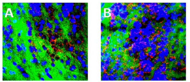
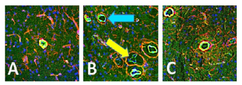
Methods: Study animals were sacrificed following endotoxin treatment and tissue sections were harvested and probed for GDNF and NOS isomers by fluorescence deconvolution microscopy. Fluorescence was mapped and quantified for each probe.
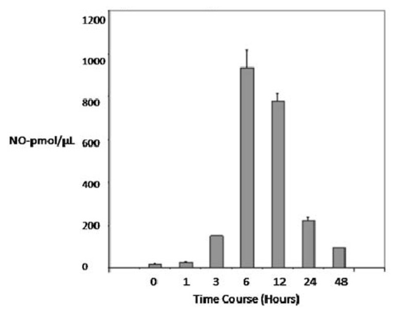
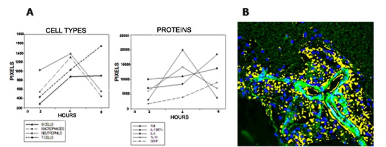
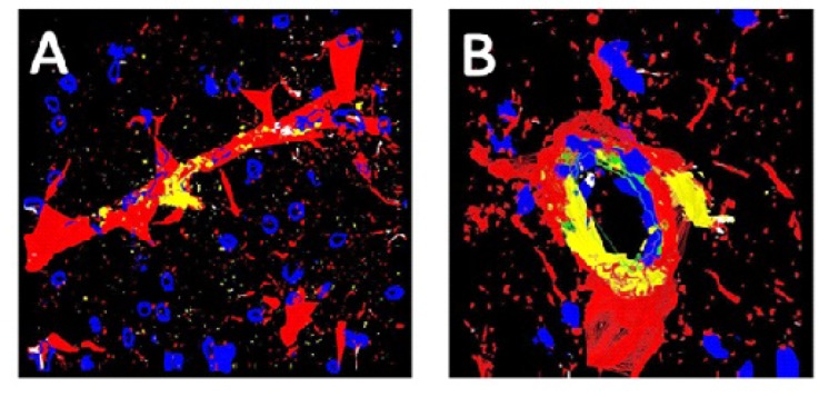
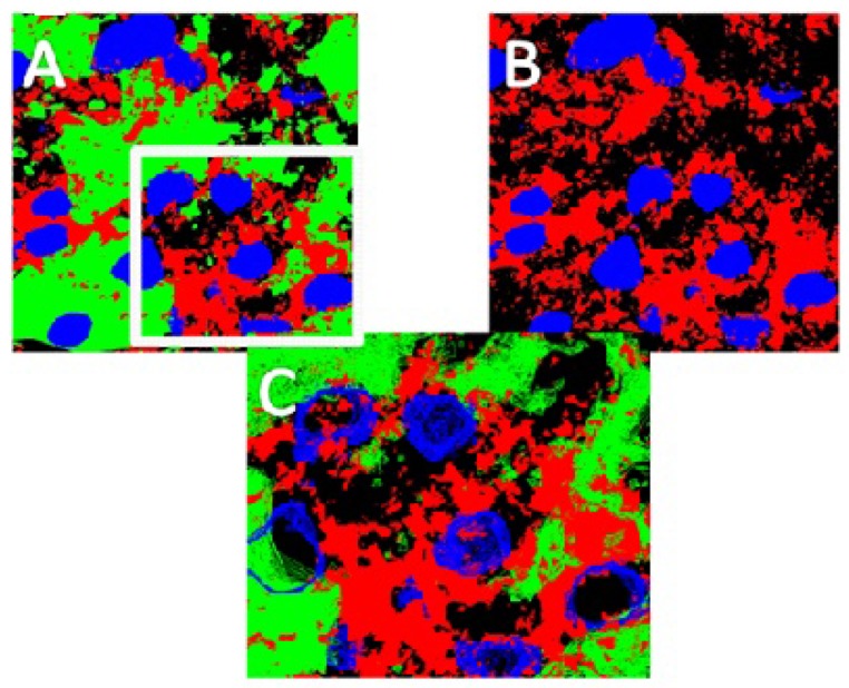
Results: An immune cell influx into 'vulnerable' areas of the brain was seen, and three NOS isomers, inducible (iNOS), neuronal (nNOS) and endothelial (eNOS), were synthesized in the brains, a finding which suggests that each isomer has a role in neurodegeneration. eNOS was found associated with blood vessels, while iNOS was associated with glial and matrix cells and nNOS was located with both glia and neurons. Following endotoxin treatment, serum levels of nitric oxide were higher at 6-8 hours, while tissue levels of NOS were elevated for much longer. Thus, induction of NOS occurred earlier than the induction of GDNF.
Conclusion: Our findings suggest that the protective abilities of GDNF to combat neural destruction are not available rapidly enough, and do not remain at sufficiently high levels long enough to assert its protective effects. (250).
Keywords: Endotoxin; Fluorescence microscopy; Glial derived neurotrophic factor; Neurodegeneration; Nitric oxide.
Figures
Similar articles
-
Normal vascular development in mice deficient in endothelial NO synthase: possible role of neuronal NO synthase.Mol Vis. 2003 Oct 8;9:549-58. Mol Vis. 2003. PMID: 14551528
-
Relative contributions of NOS isoforms during experimental colitis: endothelial-derived NOS maintains mucosal integrity.Am J Physiol Gastrointest Liver Physiol. 2004 Oct;287(4):G865-74. doi: 10.1152/ajpgi.00187.2004. Epub 2004 Jun 24. Am J Physiol Gastrointest Liver Physiol. 2004. PMID: 15217783
-
Nitric oxide in experimental joint inflammation. Benefit or detriment?Cells Tissues Organs. 2003;174(1-2):26-33. doi: 10.1159/000070572. Cells Tissues Organs. 2003. PMID: 12784039 Review.
-
Peroxynitrite and mitochondrial dysfunction in the pathogenesis of Parkinson's disease.Antioxid Redox Signal. 2003 Jun;5(3):319-35. doi: 10.1089/152308603322110896. Antioxid Redox Signal. 2003. PMID: 12880486 Review.
-
Partial cloning of constitutive and inducible nitric oxide synthases and detailed neuronal expression of NOS mRNA in the cerebellum and optic tectum of adult Atlantic salmon (Salmo salar).Brain Res Mol Brain Res. 2000 May 31;78(1-2):38-49. doi: 10.1016/s0169-328x(00)00066-8. Brain Res Mol Brain Res. 2000. PMID: 10891583
Cited by
-
Allogeneic Bone Marrow-Derived Mesenchymal Stem Cell Safety in Idiopathic Parkinson's Disease.Mov Disord. 2021 Aug;36(8):1825-1834. doi: 10.1002/mds.28582. Epub 2021 Mar 27. Mov Disord. 2021. PMID: 33772873 Free PMC article.
References
-
- Higgins G.C., Beart P.M., Shin Y.S., Chen M.J., Cheung N.S., Nagley P. Oxidative stress: emerging mitochondrial and cellular themes and variations in neuronal injury. J. Alzheimers Dis. 2010;20(Suppl. 2):S453–S473. - PubMed
LinkOut - more resources
Full Text Sources
Other Literature Sources