Optical coherence tomography based microangiography as a non-invasive imaging modality for early detection of choroido-neovascular membrane in choroidal rupture
- PMID: 27652045
- PMCID: PMC5009058
- DOI: 10.1186/s40064-016-3161-x
Optical coherence tomography based microangiography as a non-invasive imaging modality for early detection of choroido-neovascular membrane in choroidal rupture
Abstract
Introduction: To evaluate and identify early microvascular changes in patient with choroidal rupture using optical coherence tomography (OCT) based microangiography (OMAG).
Case description: One patient (one eye) with confirmed diagnosis of choroidal rupture after sustained ocular blunt trauma underwent OMAG imaging. OMAG was performed by Zeiss spectral domain OCT-angiography prototype using a "6.5 mm × 6.5 mm" field of view around macular region. The resulting images were presented into bilayers: the retinal layer and the choroidal layer.
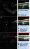
Discussion and evaluation: Choroidal rupture sites were easily shown on OMAG images with clear evidence of multiple breaks in Bruch's membrane involving macula and the region superior to nerve. OMAG provided detailed vascular network patterns in the areas of choroidal rupture, showing a concern for choroidal neovascularization (CNV). The OMAG demonstrated cross sectional area to visualize CNV location relative to the other layers of the retina, identifying functional blood vessels through the lesion. The patient's progress was followed using OMAG.
Conclusion: The images provided by OMAG give detailed microvascular findings about the macula and adjacent retinal region along with the underlying choroidal alternations. In our case, details of the architecture and vascular flow of CNVM in choroidal rupture was delivered by OMAG, which were used to follow the progression of the disease progression. Further studies are needed to assess the role of quantitative and qualitative OCT microangiography in the evaluation and treatment of choroidal rupture.
Figures


Similar articles
-
SWEPT SOURCE OPTICAL COHERENCE TOMOGRAPHY ANGIOGRAPHY OF NEOVASCULAR MACULAR TELANGIECTASIA TYPE 2.Retina. 2015 Nov;35(11):2285-99. doi: 10.1097/IAE.0000000000000840. Retina. 2015. PMID: 26457402 Free PMC article.
-
Optical coherence tomography based microangiography findings in hydroxychloroquine toxicity.Quant Imaging Med Surg. 2016 Apr;6(2):178-83. doi: 10.21037/qims.2016.01.01. Quant Imaging Med Surg. 2016. PMID: 27190770 Free PMC article.
-
Impact of intraocular pressure on changes of blood flow in the retina, choroid, and optic nerve head in rats investigated by optical microangiography.Biomed Opt Express. 2012 Sep 1;3(9):2220-33. doi: 10.1364/BOE.3.002220. Epub 2012 Aug 24. Biomed Opt Express. 2012. PMID: 23024915 Free PMC article.
-
[A new approach for studying the retinal and choroidal circulation].Nippon Ganka Gakkai Zasshi. 2004 Dec;108(12):836-61; discussion 862. Nippon Ganka Gakkai Zasshi. 2004. PMID: 15656089 Review. Japanese.
-
ZEISS Angioplex™ Spectral Domain Optical Coherence Tomography Angiography: Technical Aspects.Dev Ophthalmol. 2016;56:18-29. doi: 10.1159/000442773. Epub 2016 Mar 15. Dev Ophthalmol. 2016. PMID: 27023249 Review.
Cited by
-
A reappraisal of indirect choroidal rupture using swept-source optical coherence tomography in-vivo pathology images in patients with blunt eye trauma.Indian J Ophthalmol. 2020 Oct;68(10):2131-2135. doi: 10.4103/ijo.IJO_2192_19. Indian J Ophthalmol. 2020. PMID: 32971624 Free PMC article.
-
Optical coherence tomography angiography-guided diagnosis of a traumatic choroidal rupture-associated choroidal neovasular membrane and its management with intravitreal ranibizumab.Taiwan J Ophthalmol. 2020 Sep 22;11(3):300-304. doi: 10.4103/tjo.tjo_40_20. eCollection 2021 Jul-Sep. Taiwan J Ophthalmol. 2020. PMID: 34703748 Free PMC article.
References
-
- Huang Y, Zhang Q, Thorell MR, An L, Durbin MK, Laron M, Sharma U, Gregori G, Rosenfeld PJ, Wang RK. Swept-source OCT angiography of the retinal vasculature using intensity differentiation-based optical microangiography algorithms. Ophthalmic Surg Lasers Imaging Retin. 2014;45(5):382–389. doi: 10.3928/23258160-20140909-08. - DOI - PMC - PubMed
Grants and funding
LinkOut - more resources
Full Text Sources
Other Literature Sources

