. 2016 Sep 14:3:16060.
doi: 10.1038/mtm.2016.60. eCollection 2016.

Efficacy and biodistribution analysis of intracerebroventricular administration of an optimized scAAV9-SMN1 vector in a mouse model of spinal muscular atrophy

Affiliations

Efficacy and biodistribution analysis of intracerebroventricular administration of an optimized scAAV9-SMN1 vector in a mouse model of spinal muscular atrophy

Nicole Armbruster et al. Mol Ther Methods Clin Dev. .

Abstract

Spinal muscular atrophy (SMA) is an autosomal recessive disease of variable severity caused by mutations in the SMN1 gene. Deficiency of the ubiquitous SMN function results in spinal cord α-motor neuron degeneration and proximal muscle weakness. Gene replacement therapy with recombinant adeno-associated viral (AAV) vectors showed therapeutic efficacy in several animal models of SMA. Here, we report a study aimed at analyzing the efficacy and biodistribution of a serotype-9, self-complementary AAV vector expressing a codon-optimized human SMN1 coding sequence (coSMN1) under the control of the constitutive phosphoglycerate kinase (PGK) promoter in neonatal SMNΔ7 mice, a severe animal model of the disease. We administered the scAAV9-coSMN1 vector in the intracerebroventricular (ICV) space in a dose-escalating mode, and analyzed survival, vector biodistribution and SMN protein expression in the spinal cord and peripheral tissues. All treated mice showed a significant, dose-dependent rescue of lifespan and growth with a median survival of 346 days. Additional administration of vector by an intravenous route (ICV+IV) did not improve survival, and vector biodistribution analysis 90 days postinjection indicated that diffusion from the cerebrospinal fluid to the periphery was sufficient to rescue the SMA phenotype. These results support the preclinical development of SMN1 gene therapy by CSF vector delivery.


Figures
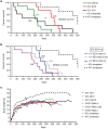
Figure 1
Figure 1
Effect of single ICV and ICV+IV combined injections of AAV9-SMN1 in SMNΔ7 mice. Kaplan-Meier survival curves (a, b): all AAV9-SMN1 doses prolonged lifespan. (a) Intracerebroventricular (ICV) injection of AAV9-SMN1 at various doses (2.5E10 vg, 4E10 vg, and 1E11 vg/mouse) in newborn SMNΔ7 mice. The median survival is given as number next to each curve (n = 8 per group). (b) Combination of ICV (2E10 vg/mouse) and intravenous (IV) vector administration at various ratios (ratio 0.25: 5E9 vg IV + 2E10 vg ICV/mouse, ratio 1: 2E10 vg IV + 2E10 vg ICV/mouse, and ratio 4: 8E10 vg IV + 2E10 vg ICV/mouse). The median survival is given as a number next to each curve (n = 8 per group). Untreated WT (n = 8) and KO mice (n = 8) were used as controls, and groups were compared using the log-rank test in a and b. (c) Body weight of untreated and treated SMNΔ7 mice with AAV9-SMN1 (ICV and ICV+IV at various doses) (n = 16 mice per group until day 90, which includes n = 8 mice/group from the biodistribution study, and thereafter “n” depending on the number of surviving mice).
Figure 2
Figure 2
AAV9-SMN1 vector biodistribution 3 months after injection. (a–e) Vector copy number (VCN), which corresponds to viral genomes/diploid genome (vg/dg), was quantified in various tissues (thoracic part of the spinal cord, liver, heart, tibialis anterior, and gastrocnemius muscles) of mutant SMNΔ7 mice 90 days post-injection. Mice received the vector at different doses by intracerebroventricular injection alone (ICV, 2.5E10 vg, 4E10 vg and 1E11 vg /mouse) and in combination with intravenous (IV) delivery at various ratios (ratio 0.25: 5E9 vg IV + 2E10 vg ICV/mouse, ratio 1: 2E10 vg IV + 2E10 vg ICV/mouse, and ratio 4: 8E10 vg IV + 2E10 vg ICV/mouse) (n = 8 for each group, with exception of the group treated by ICV at 1E11 vg/mouse, n = 3). Tissues from untreated WT (n = 8) and KO mice (n = 3) were used as negative controls (undetectable, data not shown). VCN with values <0.0005 were considered as undetectable. Spinal cord: one-way ANOVA; liver, heart, tibialis anterior, and gastrocnemius muscles: two-way analysis of variance and Bonferroni post-test; *P < 0.05, **P < 0.01, ***P < 0.001.
Figure 3
Figure 3
SMN protein localization in the spinal cord three months after vector administration in SMNΔ7 mice. (a–e) Intracerebroventricular-only delivery provided a higher amount of SMN protein in the lumbar part of the SC (shown in red, 4E10 vg/mouse). With the highest given IV dose (ratio 4) more SMN protein was detected in the heart and TA muscle. SMN levels were normalized to the loading control (α-tubulin) and the WT controls (set as 1). (f) Exemplary western blot analysis of spinal cord samples. 30 µg protein lysates were loaded (except for the muscles: 60 µg), n = 8 for each group. Spinal cord: one-way analysis of variance (ANOVA), liver, heart, tibialis anterior, and gastrocnemius muscles: two-way ANOVA and Bonferroni post-test; *P < 0.05, **P < 0.01, ***P < 0.001. (g) Immunohistochemistry for SMN protein in the anterior horn of the lumbar spinal cord. Pictures from untreated SMNΔ7 WT (left), KO (middle), and AAV9-SMN1 treated KO (right) mice are shown (AxioSCAN microscope). Scale bar, 200 µm. Insets show α-motor neurons at higher magnification.

References

    1. Crawford, TO and Pardo, CA (1996). The neurobiology of childhood spinal muscular atrophy. Neurobiol Dis 3: 97–110. - PubMed
    1. Prior, TW, Snyder, PJ, Rink, BD, Pearl, DK, Pyatt, RE, Mihal, DC et al. (2010). Newborn and carrier screening for spinal muscular atrophy. Am J Med Genet A 152A: 1608–1616. - PubMed
    1. Sugarman, EA, Nagan, N, Zhu, H, Akmaev, VR, Zhou, Z, Rohlfs, EM et al. (2012). Pan-ethnic carrier screening and prenatal diagnosis for spinal muscular atrophy: clinical laboratory analysis of >72,400 specimens. Eur J Hum Genet 20: 27–32. - PMC - PubMed
    1. Melki, J, Abdelhak, S, Sheth, P, Bachelot, MF, Burlet, P, Marcadet, A et al. (1990). Gene for chronic proximal spinal muscular atrophies maps to chromosome 5q. Nature 344: 767–768. - PubMed
    1. Lefebvre, S, Bürglen, L, Reboullet, S, Clermont, O, Burlet, P, Viollet, L et al. (1995). Identification and characterization of a spinal muscular atrophy-determining gene. Cell 80: 155–165. - PubMed