

Review
doi: 10.1615/CritRevBiomedEng.2016017198.
The Evolution of Neuroprosthetic Interfaces
Affiliations
- PMID: 27652455
- PMCID: PMC5541680
- DOI: 10.1615/CritRevBiomedEng.2016017198
Item in Clipboard
Review
The Evolution of Neuroprosthetic Interfaces
Crit Rev Biomed Eng.
2016.
Abstract
The ideal neuroprosthetic interface permits high-quality neural recording and stimulation of the nervous system while reliably providing clinical benefits over chronic periods. Although current technologies have made notable strides in this direction, significant improvements must be made to better achieve these design goals and satisfy clinical needs. This article provides an overview of the state of neuroprosthetic interfaces, starting with the design and placement of these interfaces before exploring the stimulation and recording platforms yielded from contemporary research. Finally, we outline emerging research trends in an effort to explore the potential next generation of neuroprosthetic interfaces.
Figures
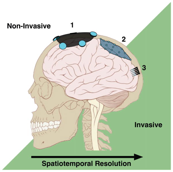
Signal resolution and NI placement. In general, the more invasive the NI the higher the accessible spatial and temporal resolution. Scalp-mounted EEG electrodes (1) and ECoG electrodes under the dura (2) record gross cortical oscillations, while intracortical electrode arrays (3) can detect single-cell activity. (Adapted from Lynch & Jaffe, 2006.)
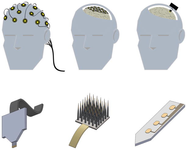
Neuroprosthetic interfaces in the CNS. Top: Placement and invasiveness for prominent BMI approaches. Current interfaces interact with the CNS at the scalp (left), the brain surface (middle), or from within the brain (right). Bottom: Examples of intracortical/penetrating neural electrodes. Intracortical NIs may take the form of microwire assemblies (left), arrays (middle), or flat shanks with multiple active recording/stimulation sites (right; center ellipses portray active sites).
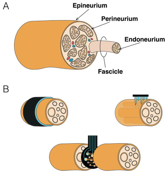
NIs in the Peripheral Nerve. (A) General overview of peripheral nerve anatomy. Individual axons are bundled in endoneurium and in turn, into discrete fascicles, supplied via blood vessels embedded in the perineurium. Fascicles in turn are bundled and protected by the dense connective tissue of the epineurium. (B) Peripheral interface electrodes range from extraneural (upper left), to penetrating/intraneural (upper right), to regenerative (bottom).
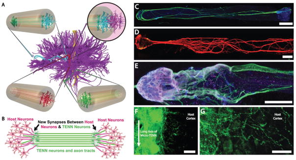
Micro-tissue engineered neural networks (Micro-TENNs), consisting of discrete neuronal population(s) with long axonal tracts within a biocompatible micro-column. Micro-TENNs are used for the direct reconstruction of long-distance axonal pathways after CNS degradation. (A) Diffusion tensor imaging representation of the human brain demonstrating the connectome comprised of long-distance axonal tracts connecting functionally distinct regions of the brain. Unidirectional (red, green) micro-TENNs and bidirectional (blue) micro-TENNs can bridge various regions of the brain (blue: corticothalamic pathway, red: nigrostriatal pathway, green: entorhinal cortex to hippocampus pathway) and synapse with host axons (purple; top right). (B) Conceptual representation of a micro-TENN forming local synapses with host neurons to form a new functional relay to replace missing or damaged axonal tracts. (C) Confocal reconstruction of a bidirectional micro-TENN, consisting of two populations of neurons spanned by long axonal tracts within a hydrogel micro-column stained via immunocytochemistry to denote axons (b-tubulin III; green), and cell nuclei (Hoechst; blue). (D) Confocal reconstruction of a unidirectional micro-TENN consisting of a single neuron population (MAP2; green) extending axons (tau; red) longitudinally (adapted from (Cullen et al., 2012)). (E) Confocal reconstruction of a unidirectional micro-TENN, stained via immunocytochemistry to denote neuronal somata/dendrites (MAP2; purple), neuronal somata/axons (tau; green) and cell nuclei (Hoechst; blue). (F) Confocal reconstruction of a transplanted GFP+ micro-TENN showing lateral outgrowth in vivo. (G) Confocal reconstruction showing GFP+ processes extending from a transplanted micro-TENN into the cortex of a rat. Scale bars: 300 μm in C, 250 μm in D, 100 μm in E, 20 μm in F and G. (Reprinted from Struzyna LA, Harris JP, Katiyar KS, Chen HI, Cullen DK, with permission from the Editorial Office of Neural Regeneration Research, Copyright 2015.)
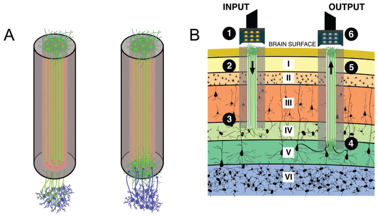
Micro-TENNs as “living electrodes.” (A) Unidirectional micro-TENNs (left) synapse with the host (blue cells) and deliver inputs to targeted cortical regions, while bidirectional micro-TENNs (right) may be synapsed by the host and transmit cortical activity. Relevant signal propagation denoted by arrows. (B) Conceptual schematic of the micro-TENNs as “living electrodes” in vivo. Left: Input paradigm. An LED array (1) optically stimulates a unidirectional micro-TENN with channelrhodopsin-positive neurons (2), which synapse with host Layer IV neurons (3). Right: Output paradigm. A microelectrode array (4) records from the neurons of a bidirectional micro-TENN (5), which are synapsed by host neurons from Layer V (6). Roman numerals denote cortical layers.
References
-
- Thurston AJ. Paré and prosthetics: the early history of artificial limbs. ANZ J Surg. 2007;77(12):1114–9. - PubMed
-
- Grill WM, Norman SE, Bellamkonda RV. Implanted neural interfaces: biochallenges and engineered solutions. Annu Rev Biomed Eng. 2009;11:1–24. - PubMed
-
- Fetz EE. Operant conditioning of cortical unit activity. Science (80-) 1969 Feb 28;163(3870):955–8. - PubMed
Publication types
MeSH terms
Grants and funding
LinkOut - more resources
Full Text Sources
Other Literature Sources