. 2016 Oct 19;27(10):2386-2399.
doi: 10.1021/acs.bioconjchem.6b00372. Epub 2016 Oct 7.

Bioorthogonal Strategy for Bioprocessing of Specific-Site-Functionalized Enveloped Influenza-Virus-Like Particles

Affiliations

Bioorthogonal Strategy for Bioprocessing of Specific-Site-Functionalized Enveloped Influenza-Virus-Like Particles

Sofia B Carvalho et al. Bioconjug Chem. .

Abstract

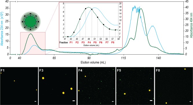
Virus-like particles (VLPs) constitute a promising platform in vaccine development and targeted drug delivery. To date, most applications use simple nonenveloped VLPs as human papillomavirus or hepatitis B vaccines, even though the envelope is known to be critical to retain the native protein folding and biological function. Here, we present tagged enveloped VLPs (TagE-VLPs) as a valuable strategy for the downstream processing and monitoring of the in vivo production of specific-site-functionalized enveloped influenza VLPs. This two-step procedure allows bioorthogonal functionalization of azide-tagged nascent influenza type A hemagglutinin proteins in the envelope of VLPs through a strain-promoted [3 + 2] alkyne-azide cycloaddition reaction. Importantly, labeling does not influence VLP production and allows for construction of functionalized VLPs without deleterious effects on their biological function. Refined discrimination and separation between VLP and baculovirus, the major impurity of the process, is achieved when this technique is combined with flow cytometry analysis, as demonstrated by atomic force microscopy. TagE-VLPs is a versatile tool broadly applicable to the production, monitoring, and purification of functionalized enveloped VLPs for vaccine design trial runs, targeted drug delivery, and molecular imaging.


Conflict of interest statement

The authors declare no competing financial interest.

Figures

Figure 1
Figure 1
Site-specific in vivo labeling of enveloped influenza VLPs. (a) Schematic representation of the procedure to metabolically introduce an azide-tagged noncanonical amino acid Aha for subsequent strain-promoted alkyne–azide [3 + 2] cycloaddition (SPAAC) labeling. During cellular protein synthesis, the Aha added to the culture medium is incorporated into nascent HA proteins. Addition of the Alexa 488-cyclooctyne reagent allows site-specific modification of HA (fluorescent tag in our case), which is reflected in VLP production. The modified HAs are incorporated into the vesicles’ envelope that is secreted from the cells that carry the chemical modification with it. (b) Confocal microscopy analysis of chemically modified VLP with the fluorescent probe Alexa 488. Dilutions (100-fold) of bulk VLPs (107 particles mL–1) were deposited onto IbiTreat 8 μ well slides. Multicolor fluorescent beads (500 nm) were used as size and green signal references (converted to grayscale). Red signal was also acquired (converted to grayscale), and from green–red merge images 500 nm beads can be discriminated from VLPs (yellow and green dots, respectively). In addition to color discrimination, for each particle detected, the full width at half-maximum was determined to evaluate the approximately 500 nm size of the control beads (red) and the subdiffraction limit VLP size (yellow) (see the Experimental section for more details). The control VLP sample shows no green signal (no labeling with Alexa 488), specific to SPAAC ligation in the experiment samples. Scale bars (white) indicate 2 μm in all images. Additional information regarding particle detection and RAW confocal images can be found in Figure S1.
Figure 2
Figure 2
Detailed interpretation of VLP polishing step by means of size-exclusion chromatography for the Alexa-488 labeled VLP. A pair of detection signals were used to monitor SEC. The elution profile was monitored by detecting the absorption of the eluted solution at both 234 nm (blue curve) and 494 nm (green curve) (emission wavelength of Alexa probe). The absorption at 234 is where roughly all biomolecules that pass through the detector contribute to the signal obtained either by absorption or by light scattering (DNA, proteins, and lipids). The detection of the absorption at 494 is specific for the fluorescent VLPs that incorporated the Alexa-488 probe. This dual detection allows better discrimination between the particles of interest (VLP) and all other contaminants such as baculoviruses. VLPs are contained in the column void volume. For each SEC fraction, confocal microscopy images were taken to monitor the presence of modified VLP (green fluorescent VLP). Scale bars (white) indicate 2 μm in all images. Images are ROI from larger independent images to better visualize the subdiffraction green dots. Merge (green–red) images are shown for clarity. According to the scheme highlighted in Figure 1, a red signal was also acquired, and from green–red merge images, 500 nm beads can be discriminated from VLPs (yellow and green dots, respectively). At the end of the SEC, between 115 and 130 mL of elution volume, concerning the elution of small molecules, there is evidence of detector signal saturation due to the elution of a high concentration of free Alexa-488 in the solution used in the labeling of VLPs.
Figure 3
Figure 3
Discrimination between VLPs and baculovirus by FACS analysis. (a) Flow cytometry of a baculovirus sample (used for infection and VLP production). A 2D correlogram of side scatter and green fluorescence signals are shown with 5% contour plots of each population. A size-scatter size ruler was made with 100, 200, and 500 nm size fluorescent beads (grayscale). Gate thresholds for negative and positive populations were performed using 100 nm bead signals. The top-right quadrant indicates green fluorescent positive >100 nm particles (VLP). In each chart, the [100–200] nm per Alexa 488 positive population gate (VLP) was built to quantify and sort the presence of labeled VLP. This analysis monitors the scatter profile of the 200–400 nm rods (red) of baculoviruses that have no green fluorescence. (b) Flow cytometry of a VLP sample before the DSP steps (blue) shows that there are clearly two particle populations: one green positive population at ≈200 nm and one with lower and nonexistent green fluorescence that has a wider size distribution. (c) Flow cytometry of F4 from the VLP SEC purification step. Analysis of the green fluorescent signal shows that the >200 nm fraction is reduced relative to A as a result of the VLP-specific green fluorescence signal. This sample was sorted with populations P1 (<200 nm population, VLP-rich) and P2 (>200 nm population, baculovirus-rich). (d) 2D correlogram of red and green fluorescence signals are shown for each population depicted in I (baculovirus), II (before DSP), and III (SEC F4). Gate thresholds for negative and positive populations were performed using 100 nm bead signals: the bottom-right quadrant is the VLP-positive quadrant (green, positive particles and red, negative particles). A significant green signal and no red signal correlates with modified VLP samples. Figures S6a,b; S7a,b; and S8a,b depict additional flow cytometry performed in the study for all steps of the DSP process.
Figure 4
Figure 4
Integrity and functionality of modified VLPs. (a) Quantification of the number of fluorescent particles detected in each DSP step in the control, unlabeled VLP, and in the labeled VLP (steps from Figure S3a). (b) Quantification of the number of fluorescent particles detected in each SEC fraction in the labeled VLP purification (SEC from Figure 2). (c) Hemagglutination assay for each step of the modified VLP purification process to assess preservation of HA biological function. (d) Hemagglutination assay for each fraction of the SEC step. (e) TEM analysis of control VLPs from the concentration step of the purification process. Scale bar indicates 100 nm. (f) TEM analysis of modified VLPs from the concentration step of the purification process. Scale bar indicates 100 nm. Uncropped and additional TEM images are available in Figure S10. The determination of the concentration of the labeled VLP solution based on the particle detection in panels c and d was also performed using eq 2 from the Experimental section and is available in Figure S4b.
Figure 5
Figure 5
Identification of HA and M1 proteins by Western blot analysis and fluorescent band detection of labeled influenza VLPs’ proteins. (a) M1 influenza protein detection on control and labeled VLPs by Western blot analysis. M1 protein from influenza A H1N1 strain was used as positive control (M1 standard). Band (1) was excised and identified as M1 by mass spectrometry. (b) HA influenza protein detection on control and labeled VLPs by Western blot analysis. H3 VLP from influenza A H3 strain was used as the positive control (H3 standard). Band (2) was excised and identified as HA by mass spectrometry. (c) SDS-PAGE gel fluorescence detection of control and labeled VLPs incubated with Alexa 488 probe. Bands (2) and (3) were excised and detected as HA by mass spectrometry. Band (4) was detected as a Telokin-like protein of baculoviruses. The term “pp” means precipitated sample.
Figure 6
Figure 6
Modified VLP detailed analysis. (a) TEM images of the major impurity in VLP production, a baculovirus, and a VLP sample before sorting revealing optimal VLP and large undesirable particles. Scale bars indicate 100 nm in all images. Uncropped and additional TEM images are available in Figure S10. (b) AFM images (error and 3D images) of a baculovirus control sample, showing rod-like morphology of this virus. Samples from each DSP step were sorted into P1 and P2, as described in Figure 3c. AFM images of P2 and P1 samples clearly show <200 nm spherical particles, consistent with VLP, and on the opposite side, the >200 nm show rod-shaped, nonspherical particles more akin to baculovirus morphology, as shown in the left AFM panels. The longitudinal (fill line) and transversal (dashed lines) cross-sections were performed to better illustrate the spherical and rod shapes of each particle visualized in each sample.

Similar articles

Cited by

References

    1. Pitoiset F.; Vazquez T.; Bellier B. (2015) Enveloped virus-like particle platforms: Vaccines of the future?. Expert Rev. Vaccines 14, 913–915. 10.1586/14760584.2015.1046440. - DOI - PubMed
    1. Plummer E. M.; Manchester M. (2011) Viral nanoparticles and virus-like particles: Platforms for contemporary vaccine design. Wires Nanomed. Nanobiotech. 3, 174–196. 10.1002/wnan.119. - DOI - PMC - PubMed
    1. Fernandes F.; Teixeira A. P.; Carinhas N.; Carrondo M. J. T; Alves P. M. (2013) Insect cells as a production platform of complex virus-like particles. Expert Rev. Vaccines 12, 225–236. 10.1586/erv.12.153. - DOI - PubMed
    1. Lua L. H. L.; Connors N. K.; Sainsbury F.; Chuan Y. P.; Wibowo N.; Middelberg A. P. J. (2014) Bioengineering virus-like particles as vaccines. Biotechnol. Bioeng. 111, 425–440. 10.1002/bit.25159. - DOI - PubMed
    1. Zhao Q. J.; Li S. W.; Yu H.; Xia N. S.; Modis Y. (2013) Virus-like particle-based human vaccines: Quality assessment based on structural and functional properties. Trends Biotechnol. 31, 654–663. 10.1016/j.tibtech.2013.09.002. - DOI - PubMed

LinkOut - more resources