. 2016 Sep 22:6:33835.
doi: 10.1038/srep33835.

Effect of graphene oxide ratio on the cell adhesion and growth behavior on a graphene oxide-coated silicon substrate

Affiliations

Effect of graphene oxide ratio on the cell adhesion and growth behavior on a graphene oxide-coated silicon substrate

Jin-Tak Jeong et al. Sci Rep. .

Abstract

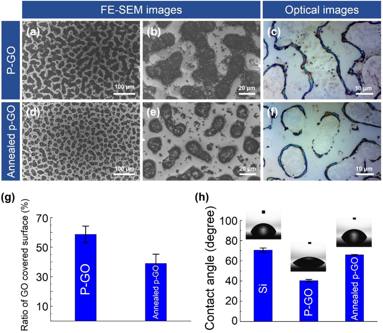
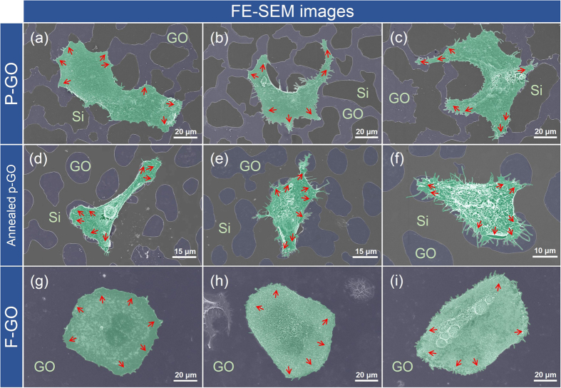
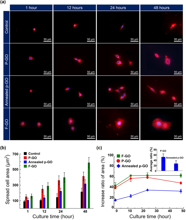
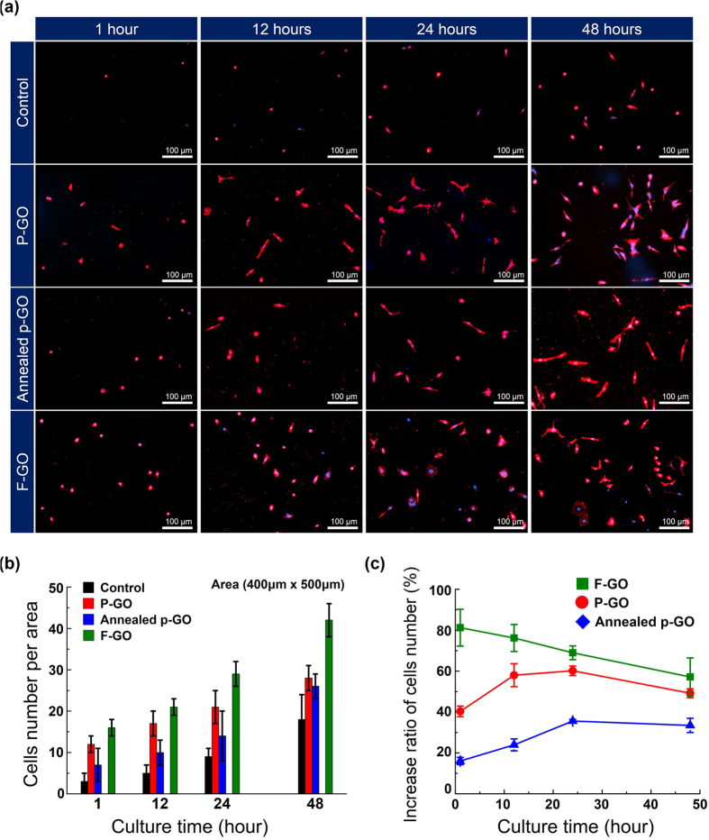
Control of living cells on biocompatible materials or on modified substrates is important for the development of bio-applications, including biosensors and implant biomaterials. The topography and hydrophobicity of substrates highly affect cell adhesion, growth, and cell growth kinetics, which is of great importance in bio-applications. Herein, we investigate the adhesion, growth, and morphology of cultured breast cancer cells on a silicon substrate, on which graphene oxides (GO) was partially formed. By minimizing the size and amount of the GO-containing solution and the further annealing process, GO-coated Si samples were prepared which partially covered the Si substrates. The coverage of GO on Si samples decreases upon annealing. The behaviors of cells cultured on two samples have been observed, i.e. partially GO-coated Si (P-GO) and annealed partially GO-coated Si (Annealed p-GO), with a different coverage of GO. Indeed, the spreading area covered by the cells and the number of cells for a given culture period in the incubator were highly dependent on the hydrophobicity and the presence of oxygenated groups on GO and Si substrates, suggesting hydrophobicity-driven cell growth. Thus, the presented method can be used to control the cell growth via an appropriate surface modification.


Figures

Figure 1
Figure 1. Schematic diagram for preparation of partially covered graphene oxide (GO) samples.
(a) and (b) Spray coating process on Si substrate using GO-containing solution. (c) Drying process and (d) annealing process of GO-coated Si samples. In (d), the partially-covered GO samples on Si substrate are prepared by minimizing the size and amount of the GO-containing solution.
Figure 2
Figure 2. Partially GO-coated Si samples.
FE-SEM images and optical images (top-view images) of (ac) P-GO and (df) Annealed p-GO samples. (g) The ratio of GO coverage and (h) contact angles on the Si surface, P-GO, and Annealed p-GO samples. In (h), the insets show the optical images of water droplets for contact angle measurement.
Figure 3
Figure 3. Raman and XPS measurements for partially GO-coated Si samples.
(a,b) Optical images and (c,d) Raman spectra of P-GO and Annealed p-GO samples. Three different positions on partially GO-coated samples, selected as Spots A, B, and C, are positioned at the inside, outside, and boundary of the thin GO films, respectively. (e,f) C1s core-level spectra of P-GO and Annealed p-GO samples.
Figure 4
Figure 4. Schematic diagram of cell culture process.
(a) Partially GO-coated Si samples (1 cm × 1 cm). (b) Sterilization process in 70% ethanol for 2 h and overnight UV exposure. (c) Cell seeding on the partially GO-coated Si samples, in which samples are covered with a PDMS well (0.8 cm in diameter). (d) Shaking the samples using a 3-D rocker for 3 min. (e) After peeling off the PDMS well, the BT-20 cells on partially GO-coated Si samples are loaded into a 24-well culture plate and incubated for 1, 12, 24, and 48 h, respectively in an incubator (37 °C, 5% CO2).
Figure 5
Figure 5. Morphology of cultured BT-20 cells on partially or fully GO-coated Si samples.
FE-SEM images (top-view images) of surface-bound BT-20 cells on (ac) P-GO, (df) Annealed p-GO, and (gi) F-GO samples. The surface-bound BT-20 cells tend to spread on the GO-coated area of the partially GO-coated samples. The red-colored arrows denote the spreading direction of surface-bound BT-20 cells on partially or fully GO-coated samples.
Figure 6
Figure 6. Spreading cell area and growth rate of cells on samples.
(a) Fluorescence images of cultured BT-20 cells on P-GO, Annealed p-GO, and F-GO samples together with the control Si sample as a function of incubation time up to 48 h. The surface-bound cells are stained by both DiI (cell membrane, red) and DAPI (cell nucleus, blue) for identifying the spreading area of the cells. (b) The average spreading areas of the surface-bound cells on these samples as a function of the incubation time up to 48 h. (c) The growth rate of cells covering the P-GO, Annealed p-GO, and F-GO samples with respect to the Si sample (control). Inset of right of (c) shows the averaged increased ratio of cell spread area for all culture times.
Figure 7
Figure 7. The number and growth rate of cells on the samples.
(a) Fluorescence images of cultured BT-20 cells on P-GO, Annealed p-GO, and F-GO samples together with the control Si sample as a function of incubation time up to 48 h. (b) The average number of cells counted by fluorescence microscopy (EVOSTM, AMG, USA) in at least 5 areas (400 × 500 μm2) on these samples. (c) The increase ratio of the average number of the cells bound on P-GO, Annealed p-GO, and F-GO samples with respect to Si samples (control).

References

    1. Wan Y. et al.. Capture, isolation and release of cancer cells with aptamer-functionalized glass bead array. Lab Chip 12, 4693–4701, 10.1039/c2lc21251j (2012). - DOI - PMC - PubMed
    1. Wan Y. et al.. Nanotextured substrates with immobilized aptamers for cancer cell isolation and cytology. Cancer 118, 1145–1154, 10.1002/cncr.26349 (2012). - DOI - PMC - PubMed
    1. Wang S. T. et al.. Highly Efficient Capture of Circulating Tumor Cells by Using Nanostructured Silicon Substrates with Integrated Chaotic Micromixers. Angew Chem Int Edit 50, 3084–3088, 10.1002/anie.201005853 (2011). - DOI - PMC - PubMed
    1. Xue P., Wu Y. F., Guo J. H. & Kang Y. J. Highly efficient capture and harvest of circulating tumor cells on a microfluidic chip integrated with herringbone and micropost arrays. Biomed Microdevices 17, 10.1007/s10544-015-9945-x (2015). - DOI - PubMed
    1. Kim S. T. et al.. Novel Streptavidin-Functionalized Silicon Nanowire Arrays for CD4(+) T Lymphocyte Separation. Nano Lett 10, 2877–2883, 10.1021/Nl100942p (2010). - DOI - PubMed

LinkOut - more resources