. 2016 Sep 22:6:33841.
doi: 10.1038/srep33841.

Pharmacological inhibitors of TRPV4 channels reduce cytokine production, restore endothelial function and increase survival in septic mice

Affiliations

Pharmacological inhibitors of TRPV4 channels reduce cytokine production, restore endothelial function and increase survival in septic mice

Thomas Dalsgaard et al. Sci Rep. .

Abstract

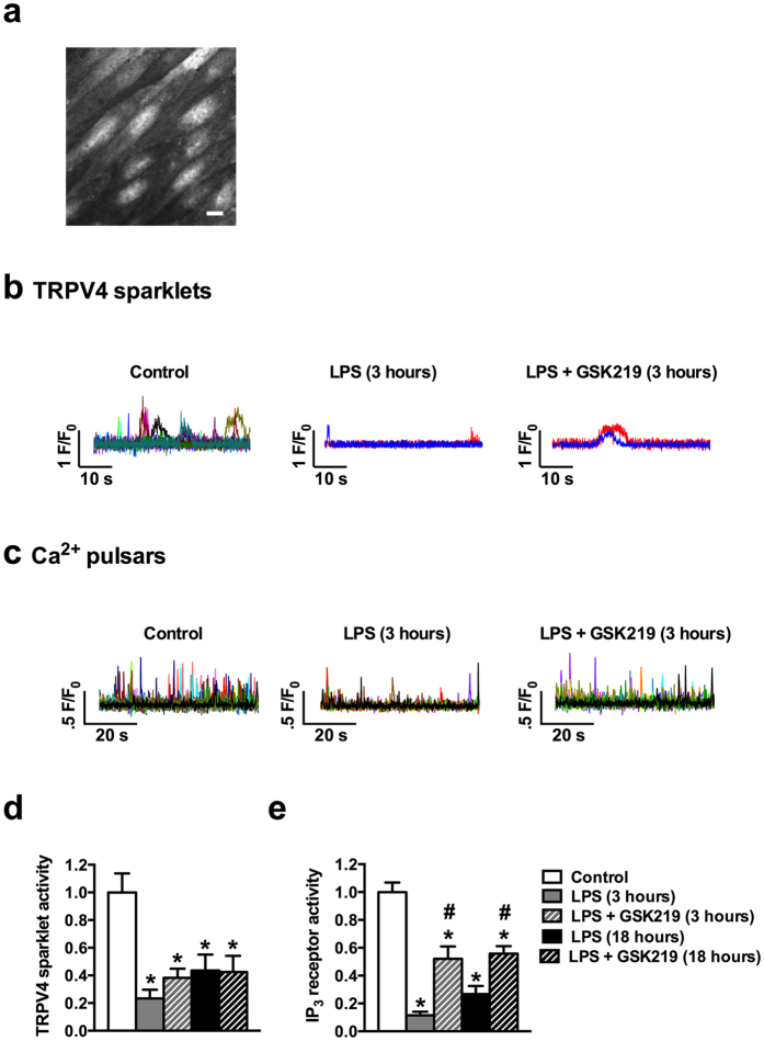
Sepsis is characterized by systemic inflammation, edema formation and hypo-perfusion leading to organ dysfunction and ultimately death. Activation of the transient receptor potential vanilloid type 4 (TRPV4) channel is associated with edema formation and circulatory collapse. Here, we show that TRPV4 channels are involved in the hyper-inflammatory response and mortality associated with sepsis. Pharmacological inhibition of TRPV4 channels in mice reduced mortality in lipopolysaccharide and cecal-ligation-and-puncture models of sepsis, but not in a tumor necrosis factor-α (TNFα)-induced sepsis model. These protective effects of TRPV4 channel inhibition were attributable to prevention of the sepsis-induced surge of a broad spectrum of pro-inflammatory cytokines, including TNFα, interleukin (IL)-1 and IL-6, and subsequent preservation of endothelial cell function, including Ca2+ signaling, integrity and endothelium-dependent vasodilation. These results suggest that TRPV4 antagonists may be of therapeutic utility in the management of sepsis.


Figures

Figure 1
Figure 1. TRPV4 channel blockade protects mice from the lethality of LPS and CLP, but not TNFα, in vivo.
(a) Seven-day survival of CLP model mice, without (CLP) or with (CLP + GSK219) GSK219; GSK219 (1 mg/kg, i.p.) was administered 1 hour prior to CLP (Sham, n = 5; CLP and CLP + GSK219, n = 12; *p < 0.05 vs. CLP alone, Mantel-Cox test). (b) Seven-day survival of mice injected with LPS only (50 mg/kg, i.p.) or LPS + GSK219; GSK219 (1 mg/kg, i.p.) was administered 1 hour prior to or 2 or 4 hours after LPS injection (n = 10 mice/group; *p < 0.05 vs. LPS treatment, Mantel-Cox test). (c) Seven-day survival of mice injected with LPS only (50 mg/kg, i.p.) or LPS + HC067; HC067 (10 mg/kg, i.p.) was administered 1 hour prior to or 2 hours after LPS injection (n = 10 mice/group; *p < 0.05 vs. LPS treatment, Mantel-Cox test). (d) Twenty-four-hour survival of mice injected with TNFα only (1 mg/kg, i.p.) or with TNFα + GSK219; GSK219 (1 mg/kg, i.p.) was administered 1 hour prior to TNFα injection (n = 10 mice/group; *p < 0.05 vs. TNFα treatment, Mantel-Cox test). (e) Seven-day survival of normal or TRPV4−/− mice injected with LPS only (50 mg/kg, i.p.) or TRPV4−/− mice injected with LPS + GSK219. GSK219 (1 mg/kg, i.p.) was administered 1 hour prior to LPS injection (n = 10 mice/group; *p < 0.05 vs. LPS treatment, Mantel-Cox test).
Figure 2
Figure 2. TRPV4 channel blockade reduces the concentration of the pro-inflammatory cytokines, TNFα, IL-1α, and IL-6, but not the anti-inflammatory cytokine, IL-10, in LPS-induced sepsis in vivo.
Blood concentrations of (a) TNFα, (b) IL-1α, (c) IL-6, and (d) IL-10, 3 and 18 hours after injection of LPS only (50 mg/kg, i.p.) or LPS + GSK219 (1 mg/kg, i.p., injected 1 hour prior to LPS). Data are expressed as means ± SEM (n = 4 mice/group; *p < 0.05 vs. control, #p < 0.05 vs. LPS treatment at the same time point, Kruskal-Wallis test with Dunn’s multiple comparisons test). IL, interleukin; TNFα, tumor necrosis factor alpha.
Figure 3
Figure 3. TRPV4 channel blockade reduces the concentration of blood markers of activated vascular endothelium in LPS-induced sepsis in vivo.
Blood concentrations of soluble (a) E-selectin, (b) ICAM-1, (c) PAI-1, (d) PECAM-1, (e) ProMMP-9, (f) P-selectin and (g) thrombomodulin, 3 and 18 hours after injection of LPS only (50 mg/kg, i.p.) or LPS + GSK219 (1 mg/kg, i.p., injected 1 hour prior to LPS). Data are expressed as means ± SEM (n = 4 mice/group; *p < 0.05 vs. control, #p < 0.05 vs. LPS treatment at the same time point, Kruskal-Wallis test with Dunn’s multiple comparisons test). ICAM, intercellular adhesion molecule; PAI, plasminogen activator inhibitor; PECAM, platelet endothelial cell adhesion molecule; MMP, matrix metallopeptidase.
Figure 4
Figure 4. TRPV4 channel blockade prevents LPS-induced endothelial dysfunction in isolated mesenteric arteries.
Vasodilation responses in isolated, pressurized (80 mmHg) mesenteric arteries from mice treated with LPS only or LPS + GSK219 measured 3 and 18 hours after LPS. In all cases, LPS and GSK219 were injected (i.p.) at doses of 50 mg/kg and 1 mg/kg, respectively; where used, GSK219 was administered 1 hour prior to LPS injection. (a–c) CCh-induced dilation in a control mouse (a), a mouse treated with LPS only, measured after 3 hours (b); a mouse treated with LPS + GSK219, measured after 3 hours (c). (d) Summary figure showing quantitative analyses of dilations to CCh (0.3–10 μM) 3 and 18 hours after treatment with LPS only or LPS + GSK219. Arteries were treated with Ca2+-free PSS at the conclusion of each experiment to obtain maximal dilation. Data are expressed as mean ± SEM (n = 5 mice/group; *p < 0.05 vs. control, #p < 0.05 vs. LPS treatment at the same time point, Kruskal-Wallis test with Dunn’s multiple comparisons test. CCh, carbachol; PSS, physiological saline solution.
Figure 5
Figure 5. TRPV4 channel blockade attenuates LPS-induced decreases in vascular endothelial TRPV4 sparklets and IP3-mediated Ca2+ pulsars.
Recordings of TRPV4 sparklets and IP3-mediated Ca2+ pulsars (measures of Ca2+-dependent endothelial cell vasoregulatory activity) in Fluo-4–loaded en face mesenteric arteries over a 60-second time period. (a) Grayscale image showing the endothelial cells in a typical field of view. Scale bar represents 10 μm. (b,c) Representative traces showing changes in Fluo-4 fluorescence caused by TRPV4 sparklets and IP3-mediated pulsars. Each colored trace represents a different region of interest from an en face preparation showing TRPV4 (b) and IP3 (c) Ca2+ events in mesenteric arteries from a control mouse; a mouse treated with LPS only, measured after 3 hours; and a mouse treated with LPS + GSK219, measured after 3 hours. In all cases, LPS and GSK219 were injected (i.p.) at doses of 50 mg/kg and 1 mg/kg, respectively; where used, GSK219 was administered 1 hour prior to LPS injection. (d,e) Summary figure showing relative TRPV4 (d) and IP3 (e) Ca2+ activity 3 and 18 hours after treatment with LPS only and LPS + GSK219. Data are expressed as mean ± SEM (n = 6 mice/group; *p < 0.05 vs. control, #p < 0.05 vs. LPS treatment at the same time point, Kruskal-Wallis test with Dunn’s multiple comparisons test). IP3, inositol trisphosphate.
Figure 6
Figure 6. TRPV4 channel blockade protects against LPS-induced increases in mesenteric artery permeability.
Pressurized (80 mmHg) mesenteric arteries, isolated from control mice and mice treated with LPS or LPS + GSK219, 3 and 18 hours after treatment, were subjected to a 40-minute pressure-drop permeability protocol, as described in Materials and Methods. In all cases, LPS and GSK219 were injected (i.p.) at doses of 50 mg/kg and 1 mg/kg, respectively; where used, GSK219 was administered 1 hour prior to LPS injection. (a) Pressure values measured as a function of time after disconnecting the pressure-servo controller and peristaltic pump. (b) Summary data of average hydraulic conductivity (Lp), a measure of endothelial permeability. Data are expressed as means ± SEM (n = 7 mice/group; *p < 0.05 vs. control, #p < 0.05 vs. LPS treatment at the same time point, Kruskal-Wallis test with Dunn’s multiple comparisons test).

References

    1. Seymour C. W. et al.. Assessment of Clinical Criteria for Sepsis: For the Third International Consensus Definitions for Sepsis and Septic Shock (Sepsis-3). JAMA: the journal of the American Medical Association 315, 762–774 (2016). - PMC - PubMed
    1. Shankar-Hari M. et al.. Developing a New Definition and Assessing New Clinical Criteria for Septic Shock: For the Third International Consensus Definitions for Sepsis and Septic Shock (Sepsis-3). JAMA: the journal of the American Medical Association 315, 775–787 (2016). - PMC - PubMed
    1. Singer M. et al.. The Third International Consensus Definitions for Sepsis and Septic Shock (Sepsis-3). JAMA: the journal of the American Medical Association 315, 801–810 (2016). - PMC - PubMed
    1. Cohen J. The immunopathogenesis of sepsis. Nature 420, 885–891 (2002). - PubMed
    1. Levi M. & van der Poll T. Inflammation and coagulation. Critical care medicine 38, S26–34 (2010). - PubMed

LinkOut - more resources