. 2016 Sep 22:5:e17716.
doi: 10.7554/eLife.17716.

The skin is a significant but overlooked anatomical reservoir for vector-borne African trypanosomes

Affiliations

The skin is a significant but overlooked anatomical reservoir for vector-borne African trypanosomes

Paul Capewell et al. Elife. .

Abstract

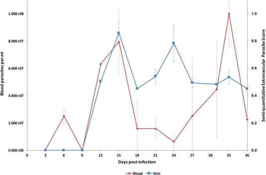
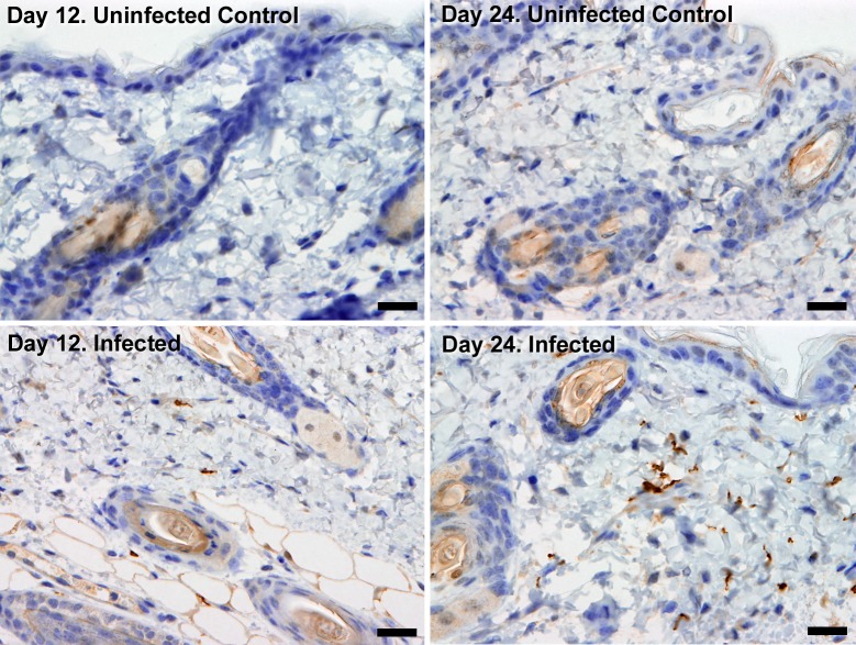
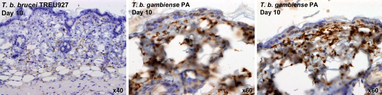
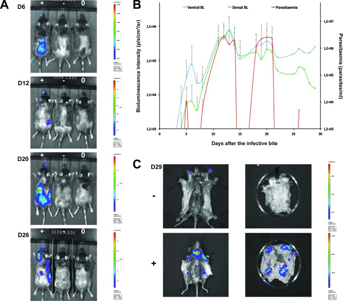
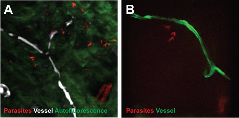
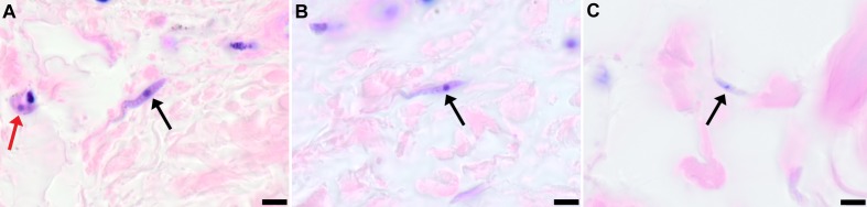
The role of mammalian skin in harbouring and transmitting arthropod-borne protozoan parasites has been overlooked for decades as these pathogens have been regarded primarily as blood-dwelling organisms. Intriguingly, infections with low or undetected blood parasites are common, particularly in the case of Human African Trypanosomiasis caused by Trypanosoma brucei gambiense. We hypothesise, therefore, the skin represents an anatomic reservoir of infection. Here we definitively show that substantial quantities of trypanosomes exist within the skin following experimental infection, which can be transmitted to the tsetse vector, even in the absence of detectable parasitaemia. Importantly, we demonstrate the presence of extravascular parasites in human skin biopsies from undiagnosed individuals. The identification of this novel reservoir requires a re-evaluation of current diagnostic methods and control policies. More broadly, our results indicate that transmission is a key evolutionary force driving parasite extravasation that could further result in tissue invasion-dependent pathology.

Keywords: Human African Trypanosomiasis; Trypanosoma brucei gambiense; epidemiology; global health; human; infectious disease; microbiology; mouse; reservoir; skin; transmission; trypanosomes.


Conflict of interest statement

The authors declare that no competing interests exist.

Figures

Figure 1.
Figure 1.. Parasite densities in the blood and in the extravascular tissue of the skin over a time-course.
The blood parasitaemia of T.b. brucei strain STIB247 (red) and the semi-quantitative score of extravascular parasites in the skin (blue) are shown over a 36-day time-course following infection in Balb/C mice. Blood parasitaemia was measured using phase microscopy using methodology outlined in (Lumsden, 1963). Skin parasite burden is an average of five high-power fields scored by histological analysis (0 = no parasites detectable; 1 = low numbers of parasites; 2 = moderate numbers of parasites; 3 = large numbers of parasites). Standard error shown (n = 3). DOI: http://dx.doi.org/10.7554/eLife.17716.002
Figure 2.
Figure 2.. Extravascular localisation of trypanosomes during an infection.
Histological sections of dorsal skin from uninfected and infected Balb/C mice stained with trypanosome-specific anti-ISG65 antibody (brown), counterstained with Gill’s Haematoxylin stain (blue) at 12 days and 24 days post-inoculation with T.b. brucei strain STIB247. Parasites are visible in extravascular locations of the skin including the deep dermis and subcutaneous adipose tissue from day 12. The scale bar represents 20 µm. DOI: http://dx.doi.org/10.7554/eLife.17716.005
Figure 2—figure supplement 1.
Figure 2—figure supplement 1.. Skin invasion by T.b. brucei strain TREU927 and T.b. gambiense strain PA.
Histological sections of dorsal skin from a mouse infected with T.b. brucei strain TREU927 at 20x magnification and two mice infected with T.b. gambiense strain PA at 40x magnification 10-days post-inoculation. Trypanosome-specific anti-ISG65 antibody reveals the presence of extravascular parasites (brown) and the slides were counterstained with Gill’s Haematoxylin stain (blue) to reveal host skin structure. DOI: http://dx.doi.org/10.7554/eLife.17716.006
Figure 3.
Figure 3.. Dynamics of parasite distribution in the extravascular tissue of the skin and in the blood during a representative course of infection following natural transmission.
A total of seven mice were infected by the single infective bite of an individual G.m. morsitans on the belly with the T.b. brucei AnTat1.1E AMLuc/tdTomato strain. Panels A and C depict representative patterns. (A) Examples of bioluminescence profiles of 3 mice (+ bitten by an infected fly, - bitten by an uninfected fly and 0 not bitten) 6, 12, 20 and 26 days after the bite are shown. (B) Ventral (blue) and dorsal (green) bioluminescence (BL) intensities (in p/s/cm2/sr on the left Y-axis) and parasitaemia (in parasites/ml in red on the right Y-axis) were measured daily for 29 days and plotted as mean ± SD (n = 7 mice). (C) The entire skins of mice (+) and (-) were dissected for bioluminescence imaging 29 days after the bite. For the mouse (+), Figure 3—figure supplement 1 shows the bioluminescence profile of dissected organs, Figure 3—figure supplement 2 presents the skin inflammation, and Figure 3—figure supplement 3 shows labelled parasites in skin sections. DOI: http://dx.doi.org/10.7554/eLife.17716.007
Figure 3—figure supplement 1.
Figure 3—figure supplement 1.. Bioluminescence mostly originates from parasites in the skin.
Mouse (+) was sacrificed and dissected for bioluminescence imaging 29 days after the infective bite. Figure 3C shows the bioluminescence profile of its entire skin and dissected organs are shown here. DOI: http://dx.doi.org/10.7554/eLife.17716.008
Figure 3—figure supplement 2.
Figure 3—figure supplement 2.. Mild inflammation of skin tissues one month after an infection by natural transmission.
After 29 days, the most bioluminescent skin region of mouse (+) was dissected, fixed in paraformaldehyde, embedded in paraffin and stained with HE. Multifocal inflammatory infiltrates containing neutrophils were located in the dermis and subcutaneous tissue and associated with oedema (arrows in A). Inflammatory foci were generally centred on blood vessels (arrows in B). DOI: http://dx.doi.org/10.7554/eLife.17716.009
Figure 3—figure supplement 3.
Figure 3—figure supplement 3.. Extravascular parasites in the skin express both VSGs and PAD1 surface markers.
After 29 days, the most bioluminescent skin region of mouse (+) was dissected, fixed in paraformaldehyde, embedded in paraffin and treated for IFA with the anti-CRD antibody that predominately labels parasites expressing VSGs (AB), or the anti-PAD1 antibody specific to transmission form 'stumpy' cells (CD). DOI: http://dx.doi.org/10.7554/eLife.17716.010
Figure 4.
Figure 4.. Extravascular localisation of trypanosomes during an infection visualised using multi-photon microscopy (A) and spinning-disk confocal microscopy (B).
(A) Still-image extracted from video (Video 1) of multi-photon live imaging of dorsal skin during a trypanosome infection. Intravenous non-targeted quantum dots (white) highlight blood vessels. T.b. brucei STIB 247 parasites transfected with mCherry to aid visualisation (red) are clearly visible and motile outside the vasculature and within the extravascular skin matrix (green). (B) Still-image extracted from (Video 3) of spinning-disk confocal live imaging of the ear of an Kdr (Flk1) C57BL/6J Rj mouse during a trypanosome infection. T.b. brucei AnTat1.1E AMLuc/tdTomato parasites expressing tdTomato (red) are moving in the extravascular region surrounding a vessel of the dermis (green). DOI: http://dx.doi.org/10.7554/eLife.17716.011
Figure 5.
Figure 5.. Extravascular localisation of trypanosomes in previously unidentified human cases of trypanosomiasis
Histological sections of skin collected from previously unidentified cases of human trypanosomiasis from the Democratic Republic of Congo, showing the presence of extravascular parasites in biopsies from three individuals (A, B and C). Skin biopsies were collected as part of a national onchocerciasis screening programme that took place in the same geographic region as an active trypanosomiasis focus. Slides were stained with Giemsa and examined under oil immersion at 100x magnification. In addition to visible slender forms (black arrows) in the extravascular tissue of the skin, a clearly identifiable stumpy transmission form with typical morphology and an unattached undulating membrane is also present in the skin of one individual (red arrow in A). The scale bar represents 5 µm. DOI: http://dx.doi.org/10.7554/eLife.17716.021

Comment in

  • Skin deep.
    Casas-Sánchez A, Acosta-Serrano Á. Casas-Sánchez A, et al. Elife. 2016 Oct 14;5:e21506. doi: 10.7554/eLife.21506. Elife. 2016. PMID: 27740910 Free PMC article.

Similar articles

Cited by

References

    1. Balyeidhusa AS, Kironde FA, Enyaru JC. Apparent lack of a domestic animal reservoir in Gambiense sleeping sickness in northwest Uganda. Veterinary Parasitology. 2012;187:157–167. doi: 10.1016/j.vetpar.2011.12.005. - DOI - PubMed
    1. Bastin P, Bagherzadeh Z, Matthews KR, Gull K. A novel epitope tag system to study protein targeting and organelle biogenesis in Trypanosoma brucei. Molecular and Biochemical Parasitology. 1996;77:235–239. doi: 10.1016/0166-6851(96)02598-4. - DOI - PubMed
    1. Berthier D, Brenière SF, Bras-Gonçalves R, Lemesre JL, Jamonneau V, Solano P, Lejon V, Thévenon S, Bucheton B. Tolerance to Trypanosomatids: A threat, or a key for disease elimination? Trends in Parasitology. 2016;32:157–168. doi: 10.1016/j.pt.2015.11.001. - DOI - PubMed
    1. Blum JA, Zellweger MJ, Burri C, Hatz C. Cardiac involvement in African and American trypanosomiasis. The Lancet Infectious Diseases. 2008;8:631–641. doi: 10.1016/S1473-3099(08)70230-5. - DOI - PubMed
    1. Branchini BR, Southworth TL, Khattak NF, Michelini E, Roda A. Red- and green-emitting firefly luciferase mutants for bioluminescent reporter applications. Analytical Biochemistry. 2005;345:140–148. doi: 10.1016/j.ab.2005.07.015. - DOI - PubMed

Publication types