Comparative Study
. 2016 Nov;89(1067):20160554.
doi: 10.1259/bjr.20160554. Epub 2016 Sep 21.

Comparison of oblique coronal images in knee of three-dimensional isotropic T2-weighted turbo spin echo MRI versus two-dimensional fast spin echo T2-weighted sequences for evaluation of posterior cruciate ligament injury

Affiliations
Comparative Study

Comparison of oblique coronal images in knee of three-dimensional isotropic T2-weighted turbo spin echo MRI versus two-dimensional fast spin echo T2-weighted sequences for evaluation of posterior cruciate ligament injury

Hee J Park et al. Br J Radiol. 2016 Nov.

Abstract

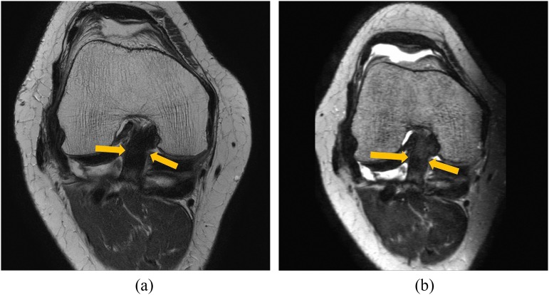
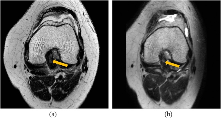
Objective: To compare image quality between three-dimensional volume isotropic turbo spin echo acquisition (3D VISTA) with the posterior cruciate ligament (PCL) view and two-dimensional (2D) fast spin echo (FSE) for evaluation of PCL injury.

Methods: This retrospective study included 60 patients with clinical suspicion of PCL injury who underwent both 2D FSE and 3D VISTA of the knee between January 2015 and December 2015. The diagnostic performance of each oblique coronal view and the combined images was evaluated for sensitivity, specificity and accuracy for diagnosing a PCL tear. The arthroscopically confirmed diagnoses were used as the reference standard. Data were analyzed using the McNemar test.

Results: The mean contrast-to-noise ratio was significantly higher for 3D VISTA than for 2D FSE. The two imaging modalities did not differ significantly in anatomical identification ability, with the exception of margin sharpness, which was inferior for 3D VISTA with Reader 2 (p = 0.038). When we classified the diagnoses of PCL injury as normal or abnormal, there were no significant differences in sensitivity, specificity or accuracy between the PCL view of 3D VISTA and 2D FSE images (p > 0.05).

Conclusion: 3D VISTA had a superior contrast-to-noise ratio than 2D FSE and similar image quality in the evaluation of the PCL. The PCL view of 3D VISTA has the same diagnostic ability as 2D FSE in the diagnosis of PCL injury and can thus replace 2D FSE. Advances in knowledge: The oblique coronal view 3D VISTA MRI has similar diagnostic ability to 2D FSE in the diagnosis of PCL injury, and therefore 3D VISTA image can replace 2D FSE.


Figures

Figure 1.
Figure 1.
(a) Sagittal T2 weighted image [repetition time/echo time (TR/TE), 4200/70] showing the technique for obtaining the oblique coronal series [posterior cruciate ligament (PCL) views]. PCL views were acquired with 2-mm slice thickness parallel to the ligament insertion site on the tibia and femur (lines in a). (b, c) The same patient as in (a). A 53-year-old female with knee pain after a slip down. The PCL view of the two-dimensional fast spin echo (TR/TE, 2500/80, b) and the PCL view image reformatted from the three-dimensional volume isotropic turbo spin echo acquisition (TR/TE, 1500/320, c) showing the PCL (arrows). Regions of interest were placed within the PCL (large circles), joint space fluid (middle circles) and air of superolateral portion of the knee joint (small circles).
Figure 2.
Figure 2.
Normal posterior cruciate ligament (PCL). A 53-year-old male with knee pain without trauma. (a) The PCL view of two-dimensional fast spin echo [repetition time/echo time (TR/TE), 2500/80] showed excellent visualization of the ligament [arrows, >75% of the entire PCL length (Score 3), >75% of the entire width of the PCL (Score 3) and excellent demarcation of the PCL from adjacent soft tissue (Score 3)]. Total anatomical identification score was 9. (b) The PCL view reformatted from three-dimensional volume isotropic turbo spin echo acquisition (TR/TE, 1500/280) showed excellent visualization of the PCL [>75% of the entire length (Score 3), >75% of the entire width of the PCL (Score 3) and excellent demarcation of the PCL from adjacent soft tissue (Score 3)]. Total anatomical identification score was 9.
Figure 3.
Figure 3.
Normal posterior cruciate ligament (PCL). A 72-year-old female with knee pain without trauma. (a) The PCL view of two-dimensional fast spin echo [repetition time/echo time (TR/TE), 2500/80] had good but not excellent visualization of the ligament [arrows, >75% of the entire PCL length (Score 3), <75% of the entire width of the PCL (Score 2) and good but not excellent demarcation of the PCL from adjacent soft tissue (Score 2)]. Total anatomical identification score was 7. (b) The PCL view reformatted from three-dimensional volume isotropic turbo spin echo acquisition (TR/TE, 1500/300) showed excellent visualization of the PCL [>75% of the entire length (Score 3), >75% of the entire width of the PCL (Score 3) and excellent demarcation of the PCL from adjacent soft tissue (Score 3)]. Total anatomical identification score was 9.
Figure 4.
Figure 4.
Partial tear of the posterior cruciate ligament (PCL). A 57-year-old female with knee pain without trauma. (a) The PCL view of two-dimensional fast spin echo [repetition time/echo time (TR/TE), 2500/80] showed a focal bright signal in the midportion of the PCL (arrow) but intact fibre continuity. (b) The PCL view reformatted from three-dimensional volume isotropic turbo spin echo acquisition (TR/TE, 1500/320) showed a focal bright signal in the midportion of the PCL (arrow) but intact fibre continuity. Partial tear of the PCL was confirmed via arthroscopy.

References

    1. Park HJ, Lee SY, Chung EC, Rho MH, Ahn JH, Kim MS, et al. . The usefulness of the oblique coronal plane in knee MRI on the evaluation of the posterior cruciate ligament. Acta Radiol 2014; 55: 961–8. doi: 10.1177/0284185113508180 - DOI - PubMed
    1. Hughston JC. The absent drawer sign in some acute posterior ligament tears of the knee. Am J Sports Med 1988; 16: 39–43. doi: 10.1177/036354658801600106 - DOI - PubMed
    1. Lysholm J, Gilquist J. Arthroscopic examination of the posterior cruciate ligament. J Bone Joint Surg Am 1981; 63: 363–6. - PubMed
    1. Yoon YC, Chung HW, Ahn JH. MR imaging of stable posterior cruciate ligament grafts in 21 arthroscopically proven cases. Korean J Radiol 2007; 8: 403–9. doi: 10.3348/kjr.2007.8.5.403 - DOI - PMC - PubMed
    1. Sherman PM, Sanders TG, Morrison WB, Schweitzer ME, Leis HT, Nusser CA. MR imaging of the posterior cruciate ligament graft: initial experience in 15 patients with clinical correlation. Radiology 2001; 221: 191–8. doi: 10.1148/radiol.2211010105 - DOI - PubMed

Publication types