. 2016 Nov 14;90(23):10629-10641.
doi: 10.1128/JVI.01222-16. Print 2016 Dec 1.

The Cytoskeletal Adaptor Obscurin-Like 1 Interacts with the Human Papillomavirus 16 (HPV16) Capsid Protein L2 and Is Required for HPV16 Endocytosis

Affiliations

The Cytoskeletal Adaptor Obscurin-Like 1 Interacts with the Human Papillomavirus 16 (HPV16) Capsid Protein L2 and Is Required for HPV16 Endocytosis

Elena Wüstenhagen et al. J Virol. .

Abstract

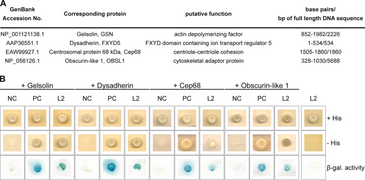
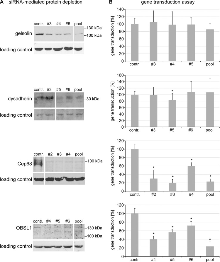
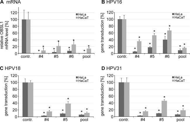
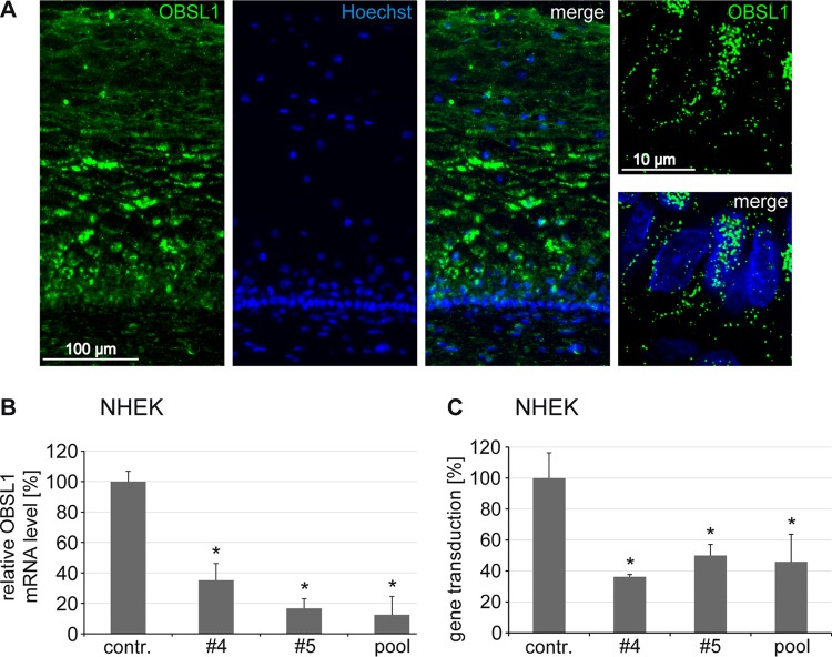
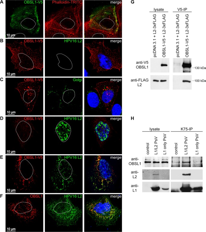
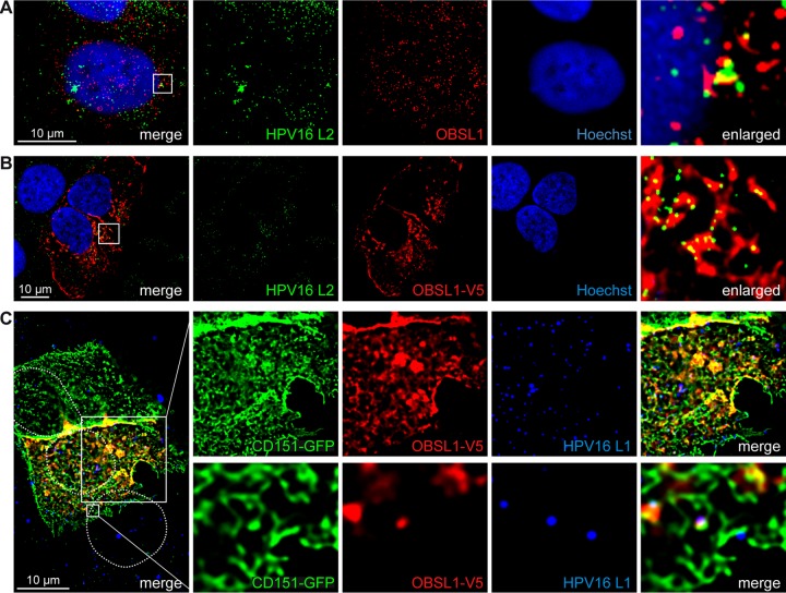
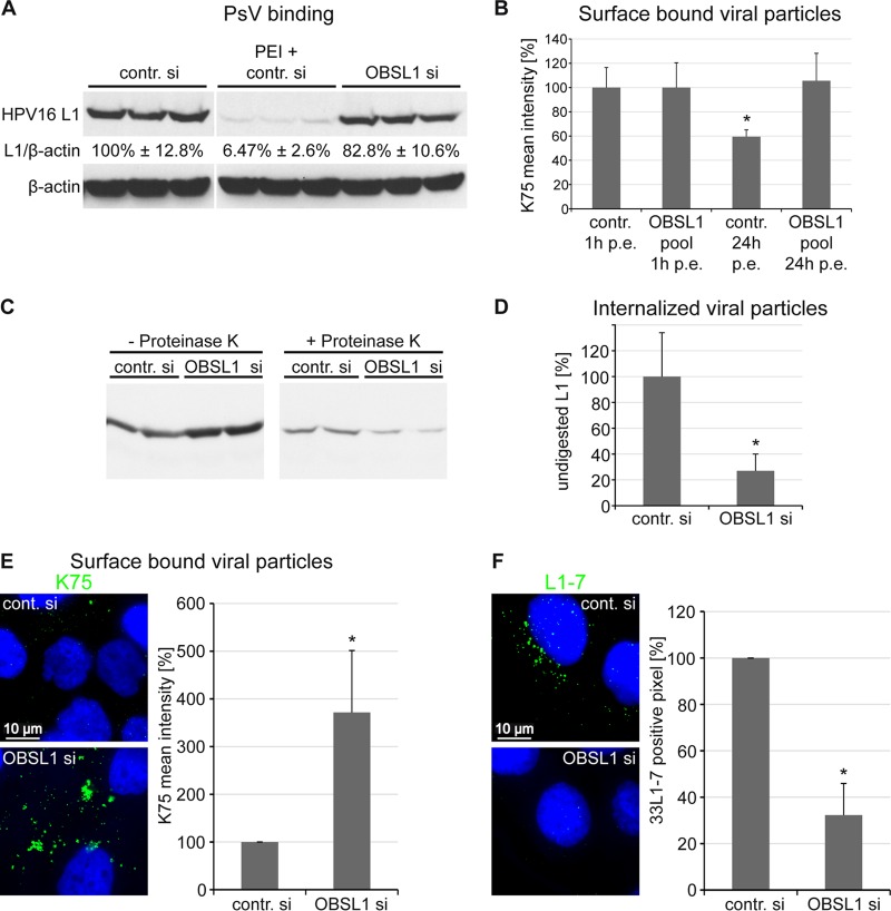
The human papillomavirus (HPV) capsid protein L2 is essential for viral entry. To gain a deeper understanding of the role of L2, we searched for novel cellular L2-interacting proteins. A yeast two-hybrid analysis uncovered the actin-depolymerizing factor gelsolin, the membrane glycoprotein dysadherin, the centrosomal protein 68 (Cep68), and the cytoskeletal adaptor protein obscurin-like 1 protein (OBSL1) as putative L2 binding molecules. Pseudovirus (PsV) infection assays identified OBSL1 as a host factor required for gene transduction by three oncogenic human papillomavirus types, HPV16, HPV18, and HPV31. In addition, we detected OBSL1 expression in cervical tissue sections and noted the involvement of OBSL1 during gene transduction of primary keratinocytes by HPV16 PsV. Complex formation of HPV16 L2 with OBSL1 was demonstrated in coimmunofluorescence and coimmunoprecipitation studies after overexpression of L2 or after PsV exposure. We observed a strong colocalization of OBSL1 with HPV16 PsV and tetraspanin CD151 at the plasma membrane, suggesting a role for OBSL1 in viral endocytosis. Indeed, viral entry assays exhibited a reduction of viral endocytosis in OBSL1-depleted cells. Our results suggest OBSL1 as a novel L2-interacting protein and endocytosis factor in HPV infection.

Importance: Human papillomaviruses infect mucosal and cutaneous epithelia, and the high-risk HPV types account for 5% of cancer cases worldwide. As recently discovered, HPV entry occurs by a clathrin-, caveolin-, and dynamin-independent endocytosis via tetraspanin-enriched microdomains. At present, the cellular proteins involved in the underlying mechanism of this type of endocytosis are under investigation. In this study, the cytoskeletal adaptor OBSL1 was discovered as a previously unrecognized interaction partner of the minor capsid protein L2 and was identified as a proviral host factor required for HPV16 endocytosis into target cells. The findings of this study advance the understanding of a so far less well-characterized endocytic pathway that is used by oncogenic HPV subtypes.


Figures

FIG 1
FIG 1
Identification of four cellular interaction partners of HPV16 L2. (A) Listed proteins were identified by yeast two-hybrid screen. The GenBank accession no., full name, putative function, and identified base pairs compared to full-length DNA sequence are listed. (B) Yeast strain L40 expressing a negative control (NC; LexA-lamin), a positive control (PC; LexA-Fos2), or the bait LexA-L2 280-473 (L2) fusion construct was transformed with corresponding prey construct B42-HA-GSN (gelsolin), B42-HA-FXYD5 (dysadherin), B42-HA-Cep68 (Cep68), B42-HA-OBSL1 (OBSL1), or empty prey vector pJG4-5 (rightmost column). Transformants were tested for the prototrophic marker histidine (− His) and β-galactosidase (β-gal.) activity reporting bait-prey interaction. Plates were incubated for 4 days at 30°C. Yeast cells were transferred onto nitrocellulose filter and incubated in 5-bromo-4-chloro-3-indolyl-β-d-galactopyranoside (X-Gal) buffer to detect β-galactosidase activity.
FIG 2
FIG 2
Influence of the putative cellular L2 interaction partners on HPV16 PsV gene transduction. HaCaT cells were transfected with control siRNA or siRNA targeted against gelsolin, dysadherin, Cep68, or OBSL1 for 48 h and then exposed to HPV16 PsVs for 24 h. (A) Cell lysates of siRNA-mediated protein depletion 48 h after transfection were analyzed by Western blotting with specific antibodies (siRNAs that showed reduction of protein level without off-target effects were selected). (B) Gene transduction efficiency was measured by luciferase activity and was normalized to lactate dehydrogenase (LDH) activity as a control for cell viability. Control siRNA-transfected cells were set to 100% ± SD. *, P < 0.05 compared to the value for the control.
FIG 3
FIG 3
OBSL1 mRNA level correlates with gene transduction by HPV16, -18, and -31 PsV in HeLa and HaCaT cells. (A) siRNA-mediated OBSL1 depletion on mRNA level in HeLa and HaCaT cells. Experiments were performed as described for Fig. 2. Forty-eight hours after siRNA transfection, total mRNA of siRNA-treated HeLa and HaCaT cells was isolated, reverse transcribed to cDNA, and analyzed by quantitative real-time PCR using gene-specific primers for amplification. Shown are the results of two independent experiments performed in duplicate. (B) HeLa and HaCaT cells were transfected with control siRNA or OBSL1 siRNA for 48 h. Shown is the gene transduction with HPV16 PsV as measured by luciferase activity and normalized to lactate dehydrogenase (LDH) activity as a control for cell viability. Control siRNA-transfected cells were set to 100% ± SD. *, P < 0.05 compared to the value for the control. (C and D) Experiments were performed as described for panel B but with exposure to HPV18 PsV (C) or HPV31 PsV (D).
FIG 4
FIG 4
Obscurin-like 1 is expressed in HPV16 target cells and required for gene transduction of primary keratinocytes. (A) Distribution of OBSL1 in cervical mucosa and expression of OBSL1 in basal cells (right images show higher magnification of basal keratinocytes). Human cervical tissue sections were stained with an OBSL1 antibody (green). Nuclei are shown in blue. (B) Normal human epidermal keratinocytes (NHEK) were transfected with control or OBSL1 siRNA for 48 h. Total mRNA was isolated, reverse transcribed to cDNA, and analyzed by quantitative real-time PCR using gene-specific primers for amplification. (C) NHEK were transfected with control or OBSL1 siRNA for 48 h and then exposed to HPV16 PsV for 24 h. Gene transduction was measured by luciferase activity and normalized by LDH measurements. The control siRNA infection rate was set to 100%. *, P < 0.05 compared to the value for the control.
FIG 5
FIG 5
HPV16 L2 forms a complex with OBSL1 in mammalian cells. (A) Confocal fluorescence microscopy shows colocalization of OBSL1-V5 with actin-binding protein phalloidin-TRITC (red). HaCaT cells were transfected with OBSL1-V5 for 24 h. OBSL1 was stained with monoclonal mouse V5 antibody (green). (B to F) Deconvolution microscopy shows OBSL1 distribution pattern. (B) Representative picture of OBSL1-V5 (red). (C) Costaining of OBSL1-V5 and the Golgi apparatus. (D) Representative nuclear HPV16 L2 distribution pattern. (E) Representative colocalization of OBSL1-V5 and L2. OBSL1-V5 and HPV16 L2 were expressed alone or in combination in HeLa cells for 24 h. OBSL1 was stained with a polyclonal rabbit V5 antibody (red), L2 with the monoclonal mouse L2 antibody L2-1 (green). The Golgi vesicles were visualized with the monoclonal mouse antibody Golgin-97 (green), and the DNA was visualized with Hoechst 33342 (blue). (F) Representative colocalization of endogenous OBSL1 and L2 in HaCaT cells. HaCaT cells were transfected with HPV16 L2 for 24 h. OBSL1 was stained using polyclonal rabbit OBSL1 antibody (red), L2 with the monoclonal mouse L2 antibody L2-1 (green), and DNA with Hoechst 33342 (blue). (G) Coimmunoprecipitation studies of L2 and OBSL1. Lysates were prepared from HeLa cells transiently cotransfected with pcDNA3.1 empty vector or OBSL1-V5 and HPV16L2-FLAG as indicated. Protein expression was verified by Western blotting of the lysates with the specific antibodies. Lysates were subjected to immunoprecipitation (IP) with antibodies specific for the V5 tag of OBSL1. The precipitated proteins were detected by Western blotting using anti-V5 or anti-FLAG antibody. (H) Coimmunoprecipitation of HPV16 PsV and OBSL1. Lysates were prepared from mock-, L1/L2 PsV-, or L1-only PsV-exposed HaCaT cells as indicated. Protein input was verified by Western blotting of the lysates with the specific antibodies. Lysates were subjected to immunoprecipitation with antibodies specific for HPV16 L1 (K75). The precipitated proteins were detected by Western blotting using anti-OBSL1 (E-16), anti-HPV16 L2 (L2-1), or anti-HPV16 L1 (312F).
FIG 6
FIG 6
OBSL1 associates with the HPV16 entry platform. (A and B) HaCaT cells were exposed to HPV16 PsV for 4 h. The cells were fixed with methanol and treated with Click-iT reaction buffer, without the addition of Alexa Fluor label for vDNA staining. Obscurin-like 1 was stained with anti-OBSL1 antibody for staining of endogenous OBSL1 (red) (A) or with anti-V5 antibody after overexpression (B), L2 was stained with monoclonal mouse anti-L2 (L2-1) (green), and nuclear DNA was stained with Hoechst 33342 (blue). (C) HaCaT cells were cotransfected with OBSL1-V5 and CD151-green fluorescent protein (GFP) for 24 h and exposed to HPV16 PsVs for 4 h. OBSL1 was stained with monoclonal mouse V5 antibody (red), L1 was stained with polyclonal rabbit L1 antibody (K75; blue), and nuclei are shown with dotted lines.
FIG 7
FIG 7
OBSL1 is involved in HPV16 internalization but not in cell surface binding. HaCaT cells were transfected for 48 h with control or OBSL1-specific siRNAs and subsequently exposed to HPV16 PsV. (A) Primary HPV16 cell surface binding is not affected by OBSL1 knockdown. siRNA-transfected cells were incubated with HPV16 PsV for 1 h at 4°C. Cell-bound PsVs were detected in cell lysates by Western blotting using the L1-specific antibody 312F (marker lane was removed). As an internal control, HaCaT cells were transfected with control siRNA and then treated with polyethyleneimine (PEI), an inhibitor of HPV16 binding. Relative band intensities were quantified densitometrically. The L1 expression level normalized to β-actin in control cells was set to 100%. (B) siRNA-transfected cells were exposed to HPV16 PsV for 1 or 24 h as indicated. The amount of surface-bound PsV was measured by flow cytometry using polyclonal L1 antibody K75. The mean fluorescence intensity of cells incubated with HPV16 PsV was adjusted to 100%. (C) siRNA-transfected cells were exposed to HPV16 PsV for 24 h. Noninternalized viral particles were removed by protease digestion (+ Proteinase K), and internalized particles were analyzed by Western blotting using the L1-specific antibody 312F. (D) Relative band intensities were quantified densitometrically: L1 level after protease digestion (+ Proteinase K) normalized to L1 input (− Proteinase K). (E) Cells were treated with siRNAs and HPV16 PsV as for panel C. The amount of surface-bound PsV was measured by quantitative fluorescence microscopy using L1 antibody K75. Representative images are shown on the left. For quantification of surface-bound viral particles, the mean K75 intensity of at least 100 cells was measured. Quantitative analysis was done using ImageJ script. The results of four independent experiments normalized to control siRNA-treated and PsV exposed cells are presented. (F) siRNA-transfected cells were exposed to HPV16 PsV for 7 h. Virus capsid disassembly was analyzed by immunofluorescence using monoclonal antibody L1-7 recognizing an epitope accessible only in internalized and disassembled capsids. Representative images are shown on the left. For quantification of viral capsid disassembly, L1-7-positive pixels of at least 100 cells were analyzed using an ImageJ script. The results of four independent experiments normalized to results for control siRNA-treated and infected cells are given. *, P < 0.05 compared to the value for the control. p.e., postexposure.

Similar articles

Cited by

References

    1. zur Hausen H. 2009. Papillomaviruses in the causation of human cancers—a brief historical account. Virology 384:260–265. doi: 10.1016/j.virol.2008.11.046. - DOI - PubMed
    1. Doorbar J, Quint W, Banks L, Bravo IG, Stoler M, Broker TR, Stanley MA. 2012. The biology and life-cycle of human papillomaviruses. Vaccine 30(Suppl 5):F55–F70. doi: 10.1016/j.vaccine.2012.06.083. - DOI - PubMed
    1. Schiller JT, Lowy DR. 2012. Understanding and learning from the success of prophylactic human papillomavirus vaccines. Nat Rev Microbiol 10:681–692. doi: 10.1038/nrmicro2872. - DOI - PMC - PubMed
    1. Baker TS, Newcomb WW, Olson NH, Cowsert LM, Olson C, Brown JC. 1991. Structures of bovine and human papillomaviruses. Analysis by cryoelectron microscopy and three-dimensional image reconstruction. Biophys J 60:1445–1456. - PMC - PubMed
    1. Buck CB, Cheng N, Thompson CD, Lowy DR, Steven AC, Schiller JT, Trus BL. 2008. Arrangement of L2 within the papillomavirus capsid. J Virol 82:5190–5197. doi: 10.1128/JVI.02726-07. - DOI - PMC - PubMed

MeSH terms

LinkOut - more resources