

Optimal echo time for functional MRI of the infant brain identified in response to noxious stimulation
- PMID: 27654315
- PMCID: PMC5516146
- DOI: 10.1002/mrm.26455
Optimal echo time for functional MRI of the infant brain identified in response to noxious stimulation
Abstract
Purpose: Blood oxygen level dependent (BOLD) brain activity, measured using functional MRI (fMRI), is dependent on the echo time (TE) and the reversible spin-spin relaxation time constant ( T2*) that describes the decay of transverse magnetization. Use of the optimal TE during fMRI experiments allows maximal sensitivity to BOLD to be achieved. Reports that T2* values are longer in infants (due to higher water concentrations and lower lipid content) have led to the use of longer TEs during infant fMRI experiments; however, the optimal TE has not been established.
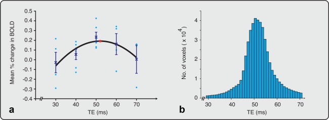
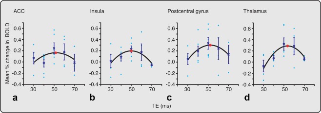
Methods: In this study, acute experimental mildly noxious stimuli were applied to the heel in 12 term infants (mean gestational age = 40 weeks, mean postnatal age = 3 days); and the percentage change in BOLD activity was calculated across a range of TEs, from 30 to 70 ms, at 3 Tesla. In addition, T2* maps of the whole brain were collected in seven infants.
Results: The maximal change in BOLD occurred at a TE of 52 ms, and the average T2* across the whole brain was 99 ms.
Conclusion: A TE of approximately 50 ms is recommended for use in 3T fMRI investigations in term infants. Magn Reson Med 78:625-631, 2017. © 2016 The Authors Magnetic Resonance in Medicine published by Wiley Periodicals, Inc. on behalf of International Society for Magnetic Resonance in Medicine.
Keywords: BOLD; T 2*; brain; echo time; imaging; infants; pain.
© 2016 The Authors Magnetic Resonance in Medicine published by Wiley Periodicals, Inc. on behalf of International Society for Magnetic Resonance in Medicine.
Figures
Similar articles
-
3D in utero quantification of T2* relaxation times in human fetal brain tissues for age optimized structural and functional MRI.Magn Reson Med. 2017 Sep;78(3):909-916. doi: 10.1002/mrm.26471. Epub 2016 Oct 3. Magn Reson Med. 2017. PMID: 27699879 Free PMC article.
-
Echo time optimization for J-difference editing of glutathione at 3T.Magn Reson Med. 2017 Feb;77(2):498-504. doi: 10.1002/mrm.26122. Epub 2016 Feb 25. Magn Reson Med. 2017. PMID: 26918659 Free PMC article.
-
Source of nonlinearity in echo-time-dependent BOLD fMRI.Magn Reson Med. 2006 Jun;55(6):1281-90. doi: 10.1002/mrm.20918. Magn Reson Med. 2006. PMID: 16700023
-
Quiet and distortion-free, whole brain BOLD fMRI using T2 -prepared RUFIS.Magn Reson Med. 2016 Apr;75(4):1402-12. doi: 10.1002/mrm.25658. Epub 2015 May 12. Magn Reson Med. 2016. PMID: 25962633
-
BOLD contrast sensitivity enhancement and artifact reduction with multiecho EPI: parallel-acquired inhomogeneity-desensitized fMRI.Magn Reson Med. 2006 Jun;55(6):1227-35. doi: 10.1002/mrm.20900. Magn Reson Med. 2006. PMID: 16680688
Cited by
-
MRI of the Neonatal Brain: A Review of Methodological Challenges and Neuroscientific Advances.J Magn Reson Imaging. 2021 May;53(5):1318-1343. doi: 10.1002/jmri.27192. Epub 2020 May 18. J Magn Reson Imaging. 2021. PMID: 32420684 Free PMC article. Review.
-
Functional neuroimaging of responses to multiple sensory stimulations in newborns with perinatal asphyxia.Transl Pediatr. 2023 Sep 18;12(9):1646-1658. doi: 10.21037/tp-23-135. Epub 2023 Sep 11. Transl Pediatr. 2023. PMID: 37814708 Free PMC article.
-
Neural correlates of gentle skin stroking in early infancy.Dev Cogn Neurosci. 2019 Feb;35:36-41. doi: 10.1016/j.dcn.2017.10.004. Epub 2017 Oct 24. Dev Cogn Neurosci. 2019. PMID: 29241822 Free PMC article.
-
Quantifying brain development in the HEALthy Brain and Child Development (HBCD) Study: The magnetic resonance imaging and spectroscopy protocol.Dev Cogn Neurosci. 2024 Dec;70:101452. doi: 10.1016/j.dcn.2024.101452. Epub 2024 Sep 21. Dev Cogn Neurosci. 2024. PMID: 39341120 Free PMC article.
-
The Significance of Echo Time in fMRI BOLD Contrast: A Clinical Study during Motor and Visual Activation Tasks at 1.5 T.Tomography. 2021 Aug 5;7(3):333-343. doi: 10.3390/tomography7030030. Tomography. 2021. PMID: 34449739 Free PMC article.
References
-
- Menon RS, Ogawa S, Tank DW, Uǧurbil K. 4 Tesla gradient recalled echo characteristics of photic stimulation‐induced signal changes in the human primary visual cortex. Magn Reson Med 1993;30:380–386. - PubMed
-
- Bandettini PA, Wong EC, Jesmanowicz A, Hinks RS, Hyde JS. Spin‐echo and gradient‐echo EPI of human brain activation using BOLD contrast: a comparative study at 1.5 T. NMR Biomed 1994;7:12–20. - PubMed
-
- van Gelderen P, Duyn JH, Liu G, Moonen CTW. Optimal T2* weighting for BOLD‐type functional MRI of the human brain. Proc Indian Acad Sci (Chem Sci) 1994;106:1617–1624.
-
- Fera F, Yongbi MN, van Gelderen P, Frank JA, Mattay VS, Duyn JH. EPI‐BOLD fMRI of human motor cortex at 1.5 T and 3.0 T: Sensitivity dependence on echo time and acquisition bandwidth. J Magn Reson Imaging 2003;19:19–26. - PubMed
-
- Wansapura JP, Holland SK, Dunn RS, Ball WS. NMR relaxation times in the human brain at 3.0 tesla. J Magn Reson Imaging 1999;9:531–538. - PubMed
Publication types
MeSH terms
Grants and funding
LinkOut - more resources
Full Text Sources
Other Literature Sources
Medical
