

Unsupervised Learning for Robust Respiratory Signal Estimation From X-Ray Fluoroscopy
- PMID: 27654320
- PMCID: PMC5489115
- DOI: 10.1109/TMI.2016.2609888
Unsupervised Learning for Robust Respiratory Signal Estimation From X-Ray Fluoroscopy
Abstract
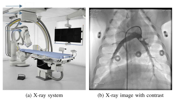
Respiratory signals are required for image gating and motion compensation in minimally invasive interventions. In X-ray fluoroscopy, extraction of a respiratory signal can be challenging due to characteristics of interventional imaging, in particular injection of contrast agent and automatic exposure control. We present a novel method for respiratory signal extraction based on dimensionality reduction that can tolerate these events. Images are divided into patches of multiple sizes. Low-dimensional embeddings are generated for each patch using illumination-invariant kernel PCA. Patches with respiratory information are selected automatically by agglomerative clustering. The signals from this respiratory cluster are combined robustly to a single respiratory signal. In the experiments, we evaluate our method on a variety of scenarios. If the diaphragm is visible, we track its superior-inferior motion as ground truth. Our method has a correlation coefficient of more than 91% with the ground truth irrespective of whether or not contrast agent injection or automatic exposure control occur. Additionally, we show that very similar signals are estimated from biplane sequences and from sequences without visible diaphragm. Since all these cases are handled automatically, the method is robust enough to be considered for use in a clinical setting.
Figures
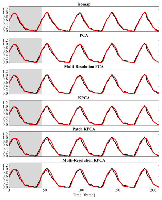
 ) are shown. For this simple sequence without contrast agent injection, all methods deliver good results. The gray background indicates the learning phase. The vertical axis is normalized to 0–1. Best viewed in color.
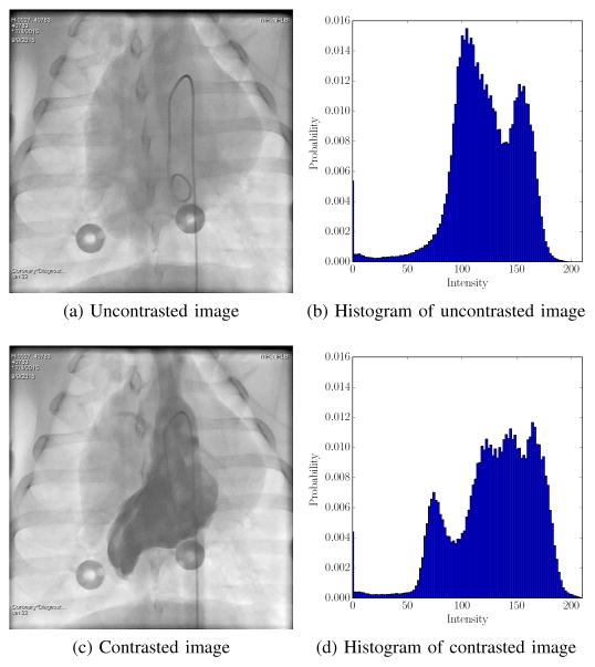
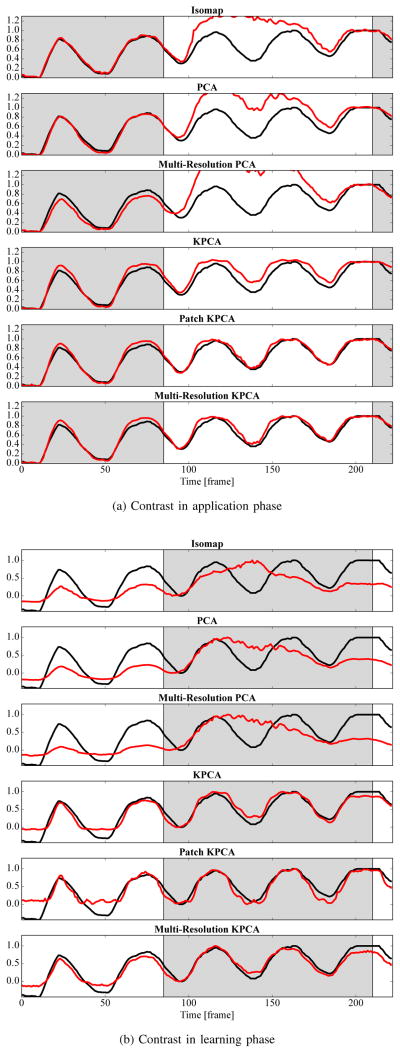 ) for an exemplary sequence with contrast agent injection. The gray background indicates the learning phase. In Fig. 6a, the contrast agent is injected in the application phase and in Fig. 6b in the learning phase. Diaphragm tracking is shown as a reference signal (–) in each plot. Best viewed in color.
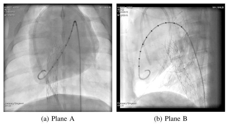
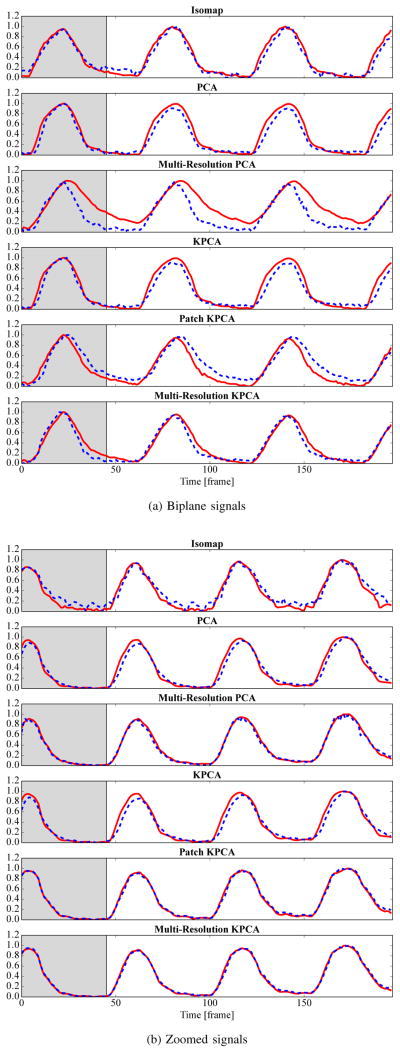 ) and plane B (
 ) are juxtaposed. In Fig. 7b, the signals are extracted from the full image (
) and a region of interest that excludes the diaphragm (
). The gray background indicates the learning phase. Best viewed in color.
) are juxtaposed. In Fig. 7b, the signals are extracted from the full image (
) and a region of interest that excludes the diaphragm (
). The gray background indicates the learning phase. Best viewed in color.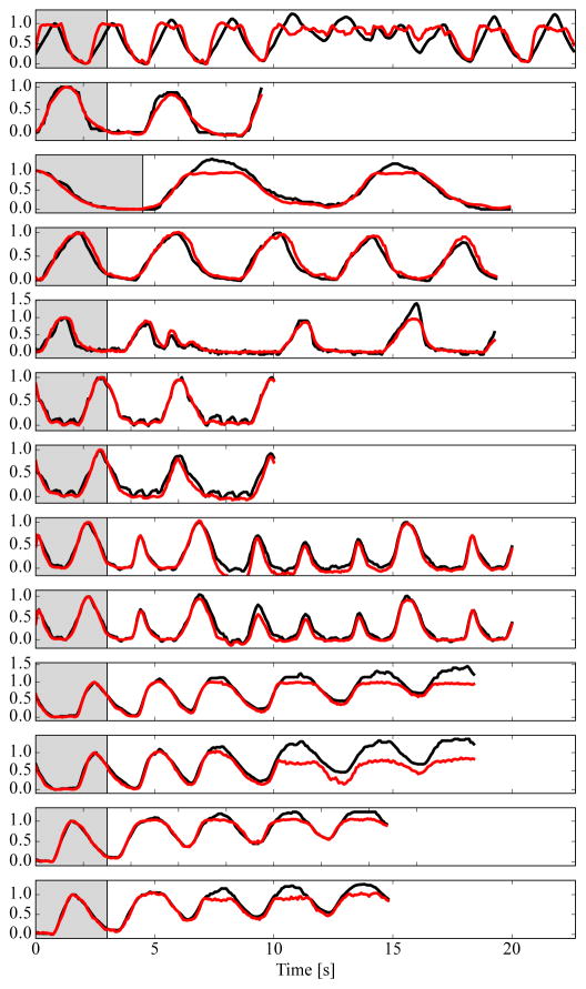 ) on free-breathing humans (top 5 sequences) and manually ventilated pigs (bottom 8 sequences). The gray background indicates the learning phase. Diaphragm tracking is shown as a reference signal (–) in each plot. Best viewed in color.
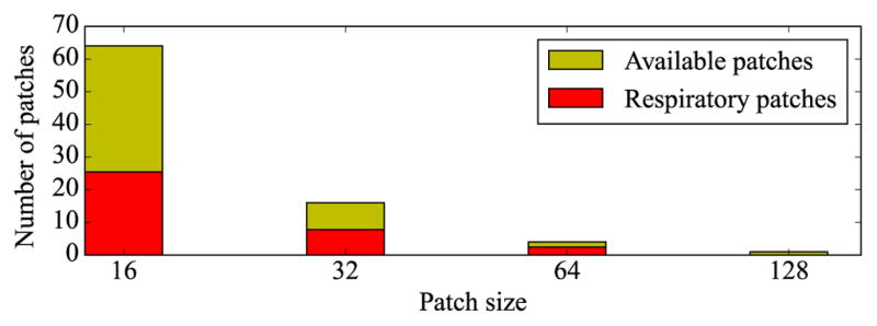
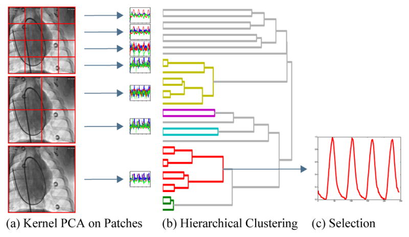
 ). For comparison, the total number of available patches in each image is given for each patch size (
 ). Best viewed in color.
 , second largest cluster
 ). On the left, the complete images are processed. On the right, a ROI without the diaphragm is used. Almost the same patches are chosen to contain respiratory information.
Similar articles
-
Motion-flow-guided recurrent network for respiratory signal estimation of x-ray angiographic image sequences.Phys Med Biol. 2020 Dec 11;65(24):245020. doi: 10.1088/1361-6560/aba087. Phys Med Biol. 2020. PMID: 32590382
-
A statistical method for retrospective cardiac and respiratory motion gating of interventional cardiac x-ray images.Med Phys. 2014 Jul;41(7):071901. doi: 10.1118/1.4881140. Med Phys. 2014. PMID: 24989379
-
A method for volumetric imaging in radiotherapy using single x-ray projection.Med Phys. 2015 May;42(5):2498-509. doi: 10.1118/1.4918577. Med Phys. 2015. PMID: 25979043 Free PMC article.
-
Image-based view-angle independent cardiorespiratory motion gating and coronary sinus catheter tracking for x-ray-guided cardiac electrophysiology procedures.Phys Med Biol. 2015 Oct 21;60(20):8087-108. doi: 10.1088/0031-9155/60/20/8087. Epub 2015 Oct 1. Phys Med Biol. 2015. PMID: 26425860
-
Verification and compensation of respiratory motion using an ultrasound imaging system.Med Phys. 2015 Mar;42(3):1193-9. doi: 10.1118/1.4907958. Med Phys. 2015. PMID: 25735274 Clinical Trial.
Cited by
-
Real-time respiratory motion compensated roadmaps for hepatic arterial interventions.Med Phys. 2021 Oct;48(10):5661-5673. doi: 10.1002/mp.15187. Epub 2021 Sep 4. Med Phys. 2021. PMID: 34431111 Free PMC article.
-
Current Research Status of Respiratory Motion for Thorax and Abdominal Treatment: A Systematic Review.Biomimetics (Basel). 2024 Mar 12;9(3):170. doi: 10.3390/biomimetics9030170. Biomimetics (Basel). 2024. PMID: 38534855 Free PMC article. Review.
-
Ensuring respiratory phase consistency to improve cardiac function quantification in real-time CMR.Magn Reson Med. 2022 Mar;87(3):1595-1604. doi: 10.1002/mrm.29064. Epub 2021 Oct 31. Magn Reson Med. 2022. PMID: 34719067 Free PMC article.
-
An MR-Based Model for Cardio-Respiratory Motion Compensation of Overlays in X-Ray Fluoroscopy.IEEE Trans Med Imaging. 2018 Jan;37(1):47-60. doi: 10.1109/TMI.2017.2723545. Epub 2017 Jul 4. IEEE Trans Med Imaging. 2018. PMID: 28692969 Free PMC article.
References
-
- Gutiérrez LF, Silva Rd, Ozturk C, Sonmez M, Stine AM, Raval AN, Raman VK, Sachdev V, Aviles RJ, Waclawiw MA, McVeigh ER, Lederman RJ. Technology preview: X-ray fused with magnetic resonance during invasive cardiovascular procedures. Catheterization and Cardiovascular Interventions. 2007;70(6):773–782. - PMC - PubMed
-
- Rhode KS, Sermesant M, Brogan D, Hegde S, Hipwell J, Lambiase P, Rosenthal E, Bucknall C, Qureshi S, Gill JS, Razavi R, Hill DL. A system for real-time XMR guided cardiovascular intervention. Medical Imaging, IEEE Transactions on. 2005;24(11):1428–1440. - PubMed
-
- King AP, Boubertakh R, Rhode KS, Ma YL, Chinchapatnam P, Gao G, Tangcharoen T, Ginks M, Cooklin M, Gill JS, Hawkes DJ, Razavi RS, Schaeffter T. A subject-specific technique for respiratory motion correction in image-guided cardiac catheterisation procedures. Medical Image Analysis. 2009;13(3):419–431. - PubMed
-
- McClelland JR, Hawkes DJ, Schaeffter T, King AP. Respiratory motion models: A review. Medical Image Analysis. 2013;17(1):19–42. - PubMed
MeSH terms
Grants and funding
LinkOut - more resources
Full Text Sources
Other Literature Sources
