. 2016 Oct;590(20):3669-3680.
doi: 10.1002/1873-3468.12427. Epub 2016 Oct 4.

Inhibition of soluble guanylyl cyclase by small molecules targeting the catalytic domain

Affiliations

Inhibition of soluble guanylyl cyclase by small molecules targeting the catalytic domain

Jagamya Vijayaraghavan et al. FEBS Lett. 2016 Oct.

Abstract

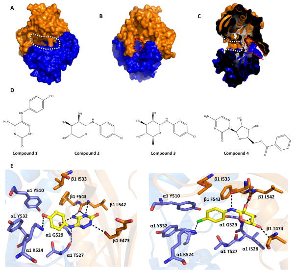
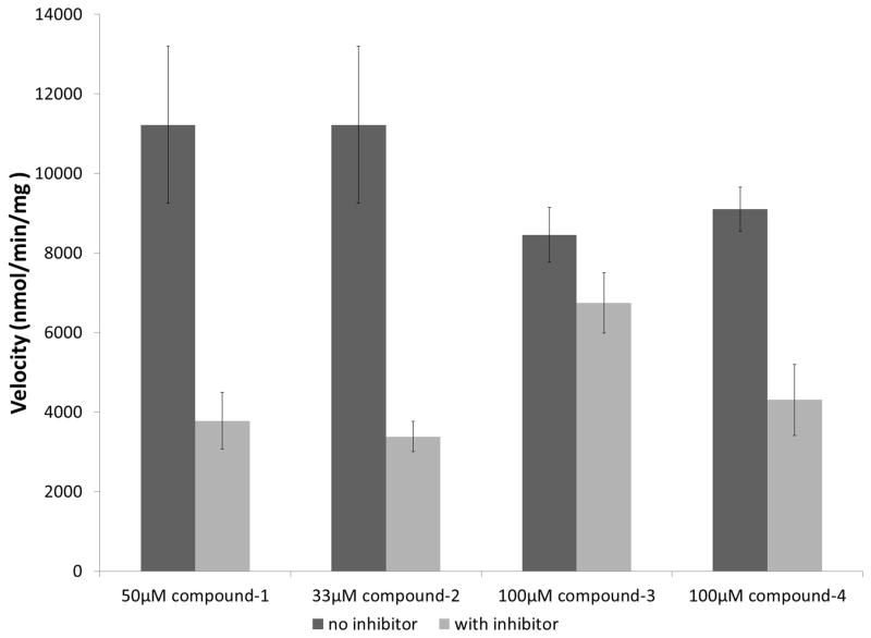
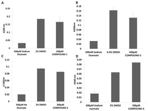
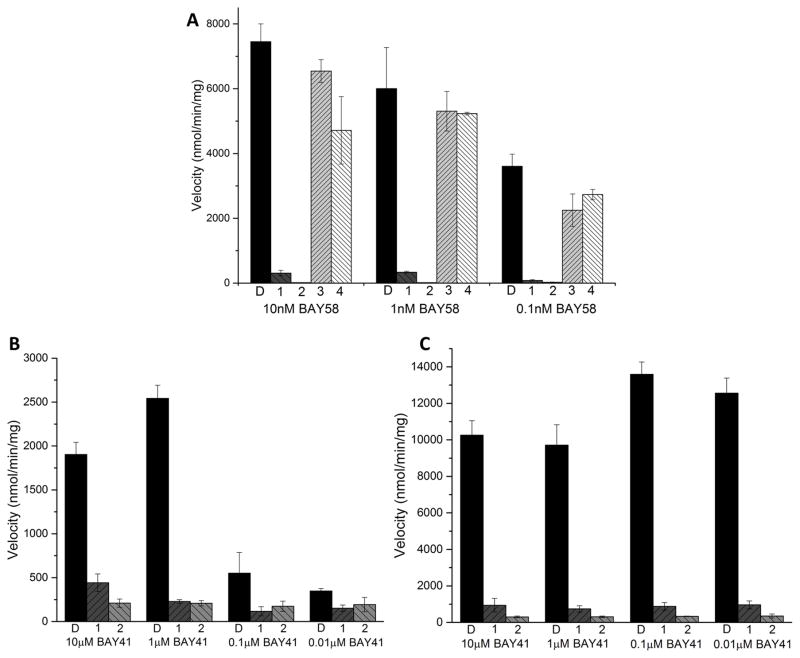
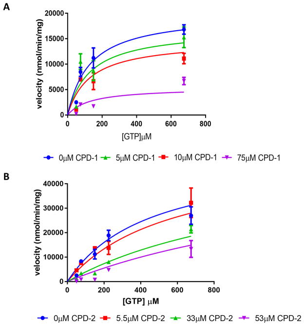
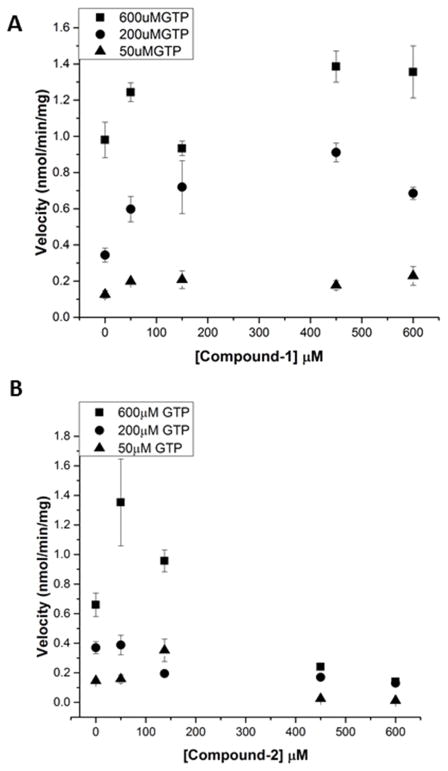
Soluble guanylyl cyclase (sGC) plays a crucial role in cyclic nucleotide signaling that regulates numerous important physiological processes. To identify new sGC inhibitors that may prevent the formation of the active catalytic domain conformation, we carried out an in silico docking screen targeting a 'backside pocket' of the inactive sGC catalytic domain structure. Compounds 1 and 2 were discovered to inhibit sGC even at high/saturating nitric oxide concentrations. Both compounds also inhibit the BAY 58-2667-activated sGC as well as BAY 41-2272-stimulated sGC activity. Additional biochemical analyses showed that compound 2 also inhibits the isolated catalytic domain, thus demonstrating functional binding to this domain. Both compounds have micromolar affinity for sGC and are potential leads to develop more potent sGC inhibitors.

Keywords: enzyme inhibition; soluble guanylyl cyclase.


Figures

Fig 1
Fig 1. In silico screening targeting the “backside pocket” of sGC catalytic domain
Molecular surface of the sGC catalytic domain showing the active site and backside pocket: A, View of the active site (outlined by white dotted line). The α1 catalytic domain is shown in blue, the β1 catalytic domain is in orange. B, Opposite face of the sGC catalytic domain showing the backside pocket (outlined by red dotted line). View in panel B is a 180° vertical rotation compared to A. C, Slabbed view of the catalytic domain dimer showing both the active site and the backside pocket. The view is obtained by a roughly 90° vertical rotation with respect to B. The active site and backside pocket are separated by a short segment of amino acids that includes residue α1 T527 (labeled as 1). D, Chemical structure of compounds 1–4. E, Predicted interactions of compound 1(left) and compound 2 (right) docking in the catalytic domain: hydrogen bonds are depicted as dashed lines. The terminal primary amine group of α K524 was not present in the structure of the catalytic domain (3UVJ), but was modelled in this figure as it can potentially form an interaction with chloride atom of compound 2. Similarly, the side chain of E473 was not included in the crystal structure but added here for illustrative purposes (this side chain is not anticipated to interact with either of the compounds). All the backside pocket residues are conserved in bovine and human sGC.
Fig 2
Fig 2. Reduction of SNAP activated sGC activity by novel small molecule inhibitors
Inhibition of SNAP (100μM) activated sGC by compounds with 150μM GTP (compounds 1 and 2) and 100μM GTP (compounds 3 and 4). The activity assays were performed as independent experiments in triplicate, error-bars represent standard error of the mean (SEM).
Fig 3
Fig 3. Analysis of promiscuous inhibition of lactate dehydrogenase
The lactate dehydrogenase (LDH) enzyme (1nM), unrelated to sGC, was used to probe for non-specific enzyme inhibition by the compounds, possibly due to promiscuous aggregation. A, LDH activity in the presence of 100μM compound 1 measured by absorbance at 500nm. B, LDH activity in the presence of 200μM compound 2. C, LDH activity in the presence of 200μM compound 3. D, LDH activity in the presence of 200μM compound 4. The inhibitor oxamate was used as a positive control as it is a known inhibitor of LDH. The DMSO bars represent uninhibited LDH with the DMSO concentration matching the DMSO concentration in the compound containing experiment. The error bars represent SEM; the experiment was done in triplicate.
Fig 4
Fig 4. Inhibition of BAY 58-2667 stimulated sGC
A, The activity of 100μM inhibitors (labeled 1–4 for compounds 1–4), or the control with 0.5% DMSO (labeled D) incubated with sGC and with varying concentrations of BAY 58-2667. B, The inhibition activity of 100μM compounds 1, 2, or 0.5% DMSO (labeled D) incubated with sGC and indicated concentrations of BAY 41-2272 in the absence of NO donor. C, The inhibitory activity of 100μM compounds 1, 2, or 0.5% DMSO (labeled D) incubated with sGC and varying concentrations of BAY 41-2272 in the presence of 1μM SNAP. The error bars represent SEM; the rate measurements were performed in triplicate.
Fig 5
Fig 5. Steady state inhibition kinetic analyses of SNAP-activated sGC
The substrate dependence of the steady state velocity of SNAP activated sGC in the presence of increasing concentrations of inhibitor. A, Compound 1. B, Compound 2. The error bars represent SEM; each point represents a rate measurement performed in triplicate. The lines represent a global fit of the data sets to a model for mixed inhibition as described in the text.
Fig 6
Fig 6. Inhibition of the sGC catalytic domain
1μM purified catalytic domain was used to determine the activity of the cGMP activity of the catalytic domain with each of the inhibitors. A, Compound 1. B, Compound 2. The error bars represent SEM; each point represents a rate measurement performed in triplicate.

References

    1. Katsuki S, Arnold W, Mittal C, Murad F. Stimulation of guanylate cyclase by sodium nitroprusside, nitroglycerin and nitric oxide in various tissue preparations and comparison to the effects of sodium azide and hydroxylamine. J Cyclic Nucleotide Res. 1977;3:23–35. - PubMed
    1. Denninger JW, Marletta MA. Guanylate cyclase and the .NO/cGMP signaling pathway. Biochim Biophys Acta. 1999;1411:334–350. - PubMed
    1. Humbert P, Niroomand F, Fischer G, Mayer B, Koesling D, Hinsch KD, Gausepohl H, Frank R, Schultz G, Bohme E. Purification of soluble guanylyl cyclase from bovine lung by a new immunoaffinity chromatographic method. Eur J Biochem. 1990;190:273–278. - PubMed
    1. Stone JR, Marletta MA. Spectral and kinetic studies on the activation of soluble guanylate cyclase by nitric oxide. Biochemistry. 1996;35:1093–1099. - PubMed
    1. Lee YC, Martin E, Murad F. Human recombinant soluble guanylyl cyclase: expression, purification, and regulation. Proc Natl Acad Sci U S A. 2000;97:10763–10768. - PMC - PubMed

MeSH terms

Substances

LinkOut - more resources