

A hypo-status in drug-dependent brain revealed by multi-modal MRI
- PMID: 27654848
- PMCID: PMC5362359
- DOI: 10.1111/adb.12459
A hypo-status in drug-dependent brain revealed by multi-modal MRI
Abstract
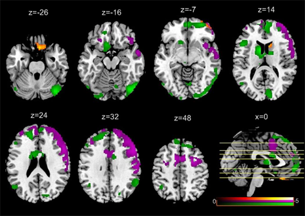
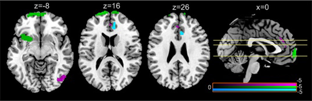
Drug addiction is a chronic brain disorder with no proven effective cure. Assessing both structural and functional brain alterations by using multi-modal, rather than purely unimodal imaging techniques, may provide a more comprehensive understanding of the brain mechanisms underlying addiction, which in turn may facilitate future treatment strategies. However, this type of research remains scarce in the literature. We acquired multi-modal magnetic resonance imaging from 20 cocaine-addicted individuals and 19 age-matched controls. Compared with controls, cocaine addicts showed a multi-modal hypo-status with (1) decreased brain tissue volume in the medial and lateral orbitofrontal cortex (OFC); (2) hypo-perfusion in the prefrontal cortex, anterior cingulate cortex, insula, right temporal cortex and dorsolateral prefrontal cortex and (3) reduced irregularity of resting state activity in the OFC and limbic areas, as well as the cingulate, visual and parietal cortices. In the cocaine-addicted brain, larger tissue volume in the medial OFC, anterior cingulate cortex and ventral striatum and smaller insular tissue volume were associated with higher cocaine dependence levels. Decreased perfusion in the amygdala and insula was also correlated with higher cocaine dependence levels. Tissue volume, perfusion, and brain entropy in the insula and prefrontal cortex, all showed a trend of negative correlation with drug craving scores. The three modalities showed voxel-wise correlation in various brain regions, and combining them improved patient versus control brain classification accuracy. These results, for the first time, demonstrate a comprehensive cocaine-dependence and craving-related hypo-status regarding the tissue volume, perfusion and resting brain irregularity in the cocaine-addicted brain.
Keywords: arterial spin labeling; brain entropy; cerebral blood flow; resting state fMRI; voxel-based morphometry.
© 2016 Society for the Study of Addiction.
Conflict of interest statement
All authors declared no conflict of interest.
Figures


Similar articles
-
Impaired functional connectivity within and between frontostriatal circuits and its association with compulsive drug use and trait impulsivity in cocaine addiction.JAMA Psychiatry. 2015 Jun;72(6):584-92. doi: 10.1001/jamapsychiatry.2015.1. JAMA Psychiatry. 2015. PMID: 25853901
-
Altered resting cerebral blood flow in adolescents with in utero cocaine exposure revealed by perfusion functional MRI.Pediatrics. 2007 Nov;120(5):e1245-54. doi: 10.1542/peds.2006-2596. Pediatrics. 2007. PMID: 17974718
-
Role of the anterior cingulate and medial orbitofrontal cortex in processing drug cues in cocaine addiction.Neuroscience. 2007 Feb 23;144(4):1153-9. doi: 10.1016/j.neuroscience.2006.11.024. Epub 2006 Dec 29. Neuroscience. 2007. PMID: 17197102 Free PMC article.
-
Brain imaging studies of cocaine abuse: implications for medication development.Crit Rev Neurobiol. 1999;13(3):227-42. doi: 10.1615/critrevneurobiol.v13.i3.10. Crit Rev Neurobiol. 1999. PMID: 10803636 Review.
-
Re-evaluating our focus in addiction: emotional dysregulation is a critical driver of relapse to drug use.Transl Psychiatry. 2024 Nov 9;14(1):467. doi: 10.1038/s41398-024-03159-5. Transl Psychiatry. 2024. PMID: 39521844 Free PMC article. Review.
Cited by
-
Cocaine addicted rats show reduced neural activity as revealed by manganese-enhanced MRI.Sci Rep. 2020 Nov 9;10(1):19353. doi: 10.1038/s41598-020-76182-3. Sci Rep. 2020. PMID: 33168866 Free PMC article.
-
Lifestyle and neurodegeneration in midlife as expressed on functional magnetic resonance imaging: A systematic review.Alzheimers Dement (N Y). 2018 May 3;4:182-194. doi: 10.1016/j.trci.2018.04.001. eCollection 2018. Alzheimers Dement (N Y). 2018. PMID: 29955662 Free PMC article. Review.
-
Multimodal Neuroimaging Differences in Nicotine Abstinent Smokers Versus Satiated Smokers.Nicotine Tob Res. 2019 May 21;21(6):755-763. doi: 10.1093/ntr/nty070. Nicotine Tob Res. 2019. PMID: 29660044 Free PMC article.
-
A Multiscale Deep Learning Method for Quantitative Visualization of Traumatic Hemoperitoneum at CT: Assessment of Feasibility and Comparison with Subjective Categorical Estimation.Radiol Artif Intell. 2020 Nov 11;2(6):e190220. doi: 10.1148/ryai.2020190220. eCollection 2020 Nov. Radiol Artif Intell. 2020. PMID: 33330848 Free PMC article.
-
rsfMRI-based brain entropy is negatively correlated with gray matter volume and surface area.Brain Struct Funct. 2025 Jan 27;230(2):35. doi: 10.1007/s00429-025-02897-6. Brain Struct Funct. 2025. PMID: 39869211
References
MeSH terms
Grants and funding
LinkOut - more resources
Full Text Sources
Other Literature Sources
Medical
