. 2016 Sep 21;14(9):e1002560.
doi: 10.1371/journal.pbio.1002560. eCollection 2016 Sep.

Dscam1 Forms a Complex with Robo1 and the N-Terminal Fragment of Slit to Promote the Growth of Longitudinal Axons

Affiliations

Dscam1 Forms a Complex with Robo1 and the N-Terminal Fragment of Slit to Promote the Growth of Longitudinal Axons

Maryam Alavi et al. PLoS Biol. .

Abstract

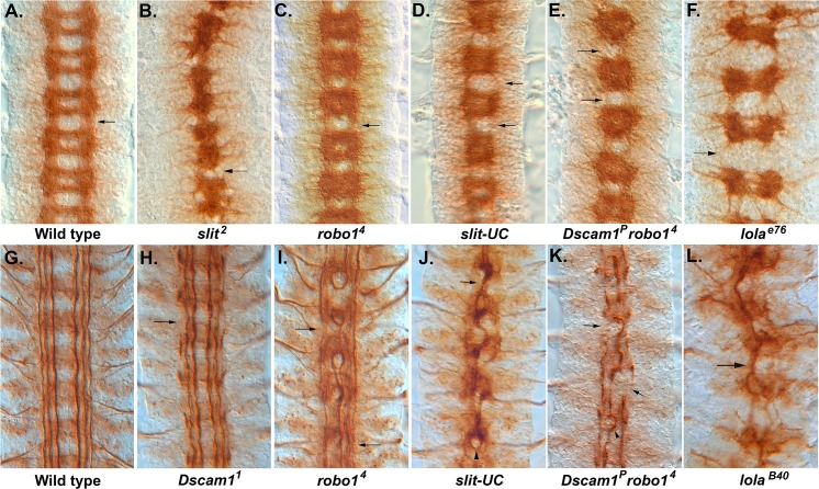
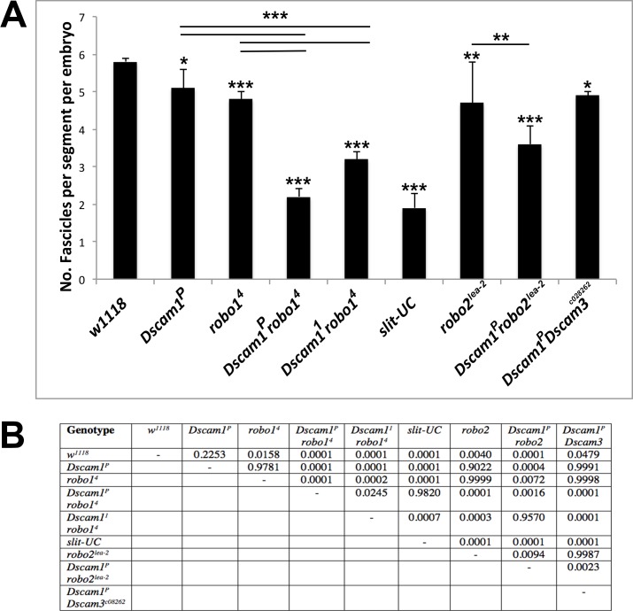
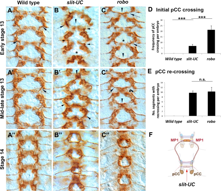
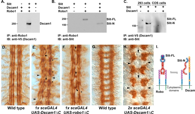
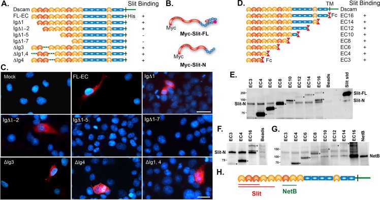
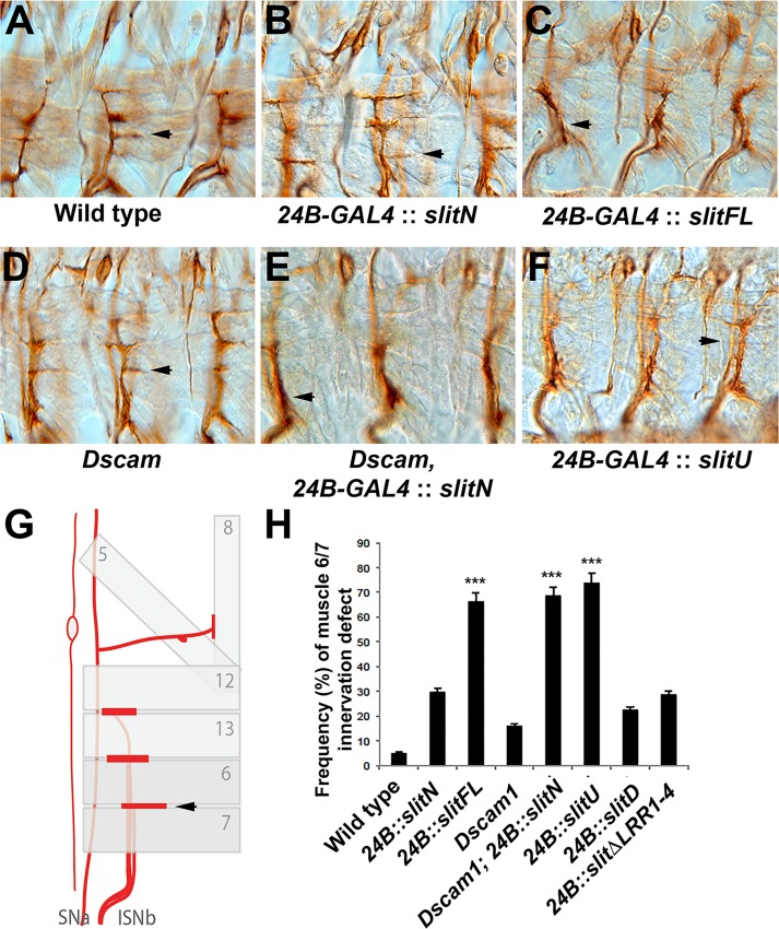
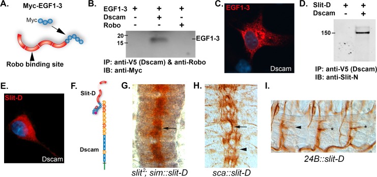
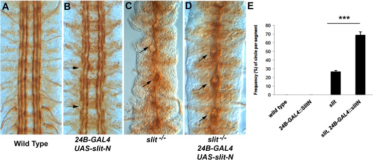
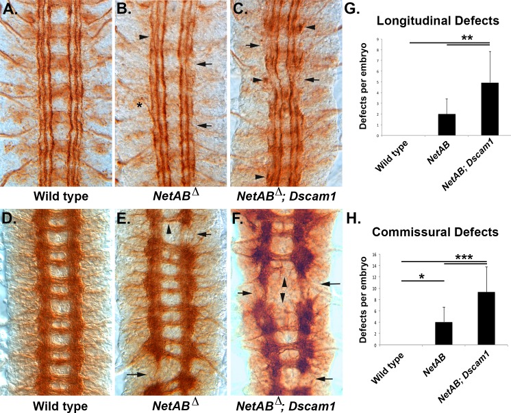
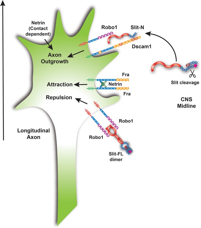
The Slit protein is a major midline repellent for central nervous system (CNS) axons. In vivo, Slit is proteolytically cleaved into N- and C-terminal fragments, but the biological significance of this is unknown. Analysis in the Drosophila ventral nerve cord of a slit allele (slit-UC) that cannot be cleaved revealed that midline repulsion is still present but longitudinal axon guidance is disrupted, particularly across segment boundaries. Double mutants for the Slit receptors Dscam1 and robo1 strongly resemble the slit-UC phenotype, suggesting they cooperate in longitudinal axon guidance, and through biochemical approaches, we found that Dscam1 and Robo1 form a complex dependent on Slit-N. In contrast, Robo1 binding alone shows a preference for full-length Slit, whereas Dscam1 only binds Slit-N. Using a variety of transgenes, we demonstrated that Dscam1 appears to modify the output of Robo/Slit complexes so that signaling is no longer repulsive. Our data suggest that the complex is promoting longitudinal axon growth across the segment boundary. The ability of Dscam1 to modify the output of other receptors in a ligand-dependent fashion may be a general principle for Dscam proteins.


Conflict of interest statement

The authors have declared that no competing interests exist.

Figures

Fig 1
Fig 1. Longitudinal axon guidance in Dscam1, robo1, and slit-UC mutants.
Embryos stained with monoclonal antibody (mAb) BP102 (A–D) to reveal the central nervous system (CNS) axon scaffold and anti-Fas2 (1D4) to reveal the longitudinal tracts (E–L). (A) Stage 16 wild-type embryo with an intersegmental longitudinal tract marked by an arrow. (B) slit2 homozygous embryo displaying the characteristic collapse of the axon scaffold onto the CNS midline. (C) robo14 homozygous embryo showing the characteristic pattern of thickened commissures and reduced longitudinals (arrow). BP102 staining in the longitudinal tracts is reduced. (D) Stage 16 uncleavable slit mutant displaying reduced or absent longitudinals (arrows) and variability in the degree of axon collapse towards the midline in each segment, e.g., a gap between the commissures is visible in the lowest segment but not in any other segment. (E) Dscam1P robo4 mutant in which several longitudinal tracts are absent (arrows). The nerve cord has not condensed as much as wild type or the robo mutant, so less segments are visible in the field of view. The individual segments are more variable than the robo mutant. (F) lolae76, a strong allele in which the longitudinal connectives (arrow) are almost completely absent. There is also a reduction in the thickness of the commissures. (G) Stage 17 wild-type embryo with longitudinal tracts running parallel to the CNS midline (center of image). (H) Dscam11 embryo with breaks to the outermost fascicle (arrow) and an overall waviness to the longitudinal fascicles. (I) robo14 mutant in which the innermost fascicles frequently merge and form circles around the midline. The medial and lateral fascicles are present (not always in the focal plane) and largely intact but display thinning and occasional breaks and defasciculation (arrows). (J) slit-UC mutant in which many of the longitudinal axons have fused to form a large fascicle that meanders along the midline (arrow). Remnants of the outermost fascicle are present but slightly out of focus, reflecting an overall degree of disorganization to the nerve cord. The occasional robo-like circle is visible (arrowhead). (K) Dscam1P robo4 homozygous embryo displaying disruption to all the longitudinal axon tracts. A large, thick fascicle follows the midline, occasionally forming circles (arrowhead). The outermost fascicle is still present but is thicker than normal, so it has likely fused with the intermediate fascicle. There are frequent breaks, and sometimes the fascicle is completely absent (arrows). (L) lolaB40 strong allele showing a large fascicle wandering back and forth across the midline (arrow). The underlying data are shown in S1 Data.
Fig 2
Fig 2. Statistical analysis of longitudinal axon guidance phenotypes.
For each of the mutant genotypes shown, the number of fascicles visible at the segment boundaries was counted in stage 17 embryos stained with anti-Fas2. The number of fascicles was divided by the number of segments scored in the embryo to get an average number of fascicles per segment, which is six in wild type (S1 Data). w1118 was used as wild type. (A) Graph displaying the results of quantification. The data were analyzed by using a Tukey honest significant difference (HSD) within a one-way ANOVA. Statistical significant differences relative to wild type are shown above each individual column (* p < 0.05, ** p < 0.01, *** p < 0.0001). Select pairwise comparisons are shown as horizontal bars. Error bars are one standard deviation. (B) Complete results of the ANOVA showing the significance of all pairwise comparisons. The underlying data are shown in S1 Data.
Fig 3
Fig 3. Analysis of longitudinal pioneer axons in an uncleavable slit mutant.
Anti-Fasciclin 2 (mAb 1D4) staining of embryos during early CNS development (stages 13–14 as indicated). The anterior is to the top of the image. (A) Wild-type embryo in which the pCC growth cones (arrows) are growing anteriorly and away from the midline. (B) A slit-UC mutant in which the pCC growth cones (arrows) are growing anteriorly and away from the midline (arrowhead), resembling wild-type behavior. However, the growth cones are growing at different rates. The cell bodies are also positioned closer to the midline, particularly in the lowest segment (asterisk). (C) robo4 mutant in which the pCC growth cones (arrows) have grown towards the midline and fasciculated with their contralateral homologues (arrowheads). The asterisk marks a relatively rare occurrence in which the pCC axons failed to cross the midline. (A′) Mid-stage 13 wild-type embryo in which the pCC axons have met and fasciculated with descending longitudinal axon pioneers. (B′) slit-UC embryo in which most pCC axons have continuing growing ipsilaterally towards descending longitudinal axon pioneers (arrows), but in one segment, the pCC axons have grown towards the midline and fasciculated (arrowhead). Although the axon behavior is similar to wild type, the cell body positioning is irregular and more closely resembles robo. (C′) robo mutant in which the pCC axons (arrows) have crossed the midline (arrowhead) and are growing towards the descending pioneer axons. The axons are beginning to form the characteristic circular fascicles of the robo mutant. (A′′) Wild-type embryo in which the pioneer longitudinal tracts have continued their growth on either side of the midline but do not cross it. (B′′) slit-UC mutant in which the longitudinal pioneers, both ascending and descending, have chosen to cross the CNS midline rather than continue growing parallel to the CNS midline. The 1D4-positive fascicles have formed the characteristic circles of the robo phenotype. The positioning of cell bodies and the size of the circles formed by the pioneers continue to be irregular. (C′′) robo mutant in which the circular fascicles created by longitudinal pioneers recrossing the midline in every segment are visible. (D) Quantification of initial pCC midline crossing in early to mid-stage 13 embryos. The number of individual pCC axons inappropriately crossing the midline was scored for the 8 abdominal and 3 thoracic segments for individual embryos (total of 22 pCC axons; S2 Data). Genotypes are shown beneath the columns. Error bars are one standard deviation. The data were analyzed using a Tukey HSD within a one-way ANOVA. Horizontal bars indicate pairwise comparisons, and there is a statistically significant difference between robo mutants and slit-UC (*** p < 0.0001). (E) The crossing of pCC axons at the second commissure encountered was analyzed in the abdominal and thoracic segments of stage 14 embryos (11 segments; S3 Data). The presence of a pCC axon at or near the midline was scored as a crossing event. Genotypes are shown beneath the columns, and error bars are one standard deviation. The data were analyzed using a Tukey HSD within a one-way ANOVA. There was no significant difference between robo and slit-UC mutants. (F) Schematic representing the axon trajectories observed in slit-UC mutants. The pCC axons are born just posterior to a commissure (red arrow). Ninety-six percent of robo mutant pCC axons immediately cross this commissure, but only 30% of slit-UC mutant axons do. The pCC axons grow anteriorly, encountering the descending MP1 longitudinal pioneer axons. In both robo and slit-UC mutants, the pCC axon typically follows the MP1 axon to the midline. The primary difference between robo and slit-UC mutants is therefore the tendency to cross the midline at the first commissure (red arrow). The MP1 axon also inappropriately crosses the midline on encountering a commissure. For the full schematic, see S3 Fig. The underlying data are shown in S2 Data and S3 Data.
Fig 4
Fig 4. Dscam1 forms a Slit-N-dependent complex with Robo1.
(A) Transfection of COS-7 and 293 cells with plasmids encoding Dscam1, Robo1, and Slit as indicated by pluses (presence of plasmid) above each lane. Immunoprecipitation with an anti-Robo1 antibody pulls down Dscam1, but only in the presence of Slit. (B) Immunoprecipitation of Robo1 alone recovers full-length Slit, but in the presence of Dscam1, Slit-N is preferentially recovered. (C) Schematic summarizing the observed interactions, suggesting that an equilibrium between Robo1 homodimers and Dscam1-Robo1 heterodimers exists. Slit-FL is depicted as dimerizing at the leucine-rich repeat 4 (LRR4) and cysteine knot domains. The stoichiometry of the Dscam1-Robo1-Slit-N complex is unknown. (D) Anti-Fasciclin 2 (MAb 1D4) staining of longitudinal fascicles. Three tracts project in an anterior-posterior direction on either side of the CNS midline, which is in the center of the picture. (E) A single copy (represented by “1x” in the figure legend) of a Dscam1ΔC transgene expressed pan-neurally by the scabrous promoter (sca-GAL4) reveals severe disruption of the longitudinal fascicles. In addition to the overall disorganization of the fascicles, longitudinal breaks (arrows) can also be seen, as well as clumping and stalling (asterisks). There is midline crossing at only one point (arrow). (F) A single copy of a roboΔC transgene expressed pan-neurally by the scabrous promoter reveals midline crossing of the innermost fascicle at multiple points (arrowheads). The longitudinal tracts have increased waviness and are sometimes merged or absent (asterisk), but they are generally continuous. (G) BP102 staining to reveal the wild-type CNS axon scaffold with its characteristic ladder-like pattern. (H) Multiple copies (most likely two of each, represented by “2x” in the legend) of a Dscam1ΔC transgene and sca-GAL4 driver produce absence of the longitudinal connectives between segments (arrowheads) and greatly reduced midline crossing (arrows). (I) Model of Robo binding Slit-FL as a homodimer and Slit-N binding Dscam and Robo as a heterodimer; the stoichiometry of the complex is unknown at present and could involve multiple molecules. The relationship between the two complexes is shown as an equilibrium that could be altered by overexpression or removal of specific components.
Fig 5
Fig 5. Mapping of Slit-N and Netrin-B binding sites in Dscam1.
(A) Schematic representation of the Dscam1 expression constructs used. Ig domains are either orange for constant domains or red for variable domains, and fibronectin (FN) domains are blue rectangles. The naming scheme for the constructs indicates which Ig domains are deleted (Δ), while the construct with the entire ectodomain is called FL-EC. All constructs had their cytoplasmic domain replaced with a 6xHis epitope tag. Immunoblot with an anti-His antibody indicates that all constructs are expressed at their predicted sizes. (B) Schematic of N-terminally Myc epitope-tagged Slit constructs generated with leucine-rich repeats (LRRs) in red, epidermal growth factor (EGF) repeats in blue, the Laminin G domain in bright pink, and the cysteine knot in dull pink. (C) Cell overlay assays. COS cells were transfected with different Dscam1 expression constructs and tested for binding to N-terminally myc-tagged Slit-N produced by 293 cells. The relevant constructs are indicated. Binding of myc-Slit-N was detected by anti-myc (red) and counterstained with 4′,6-diamidino-2-phenylindole (DAPI) to reveal the nuclei (blue). Myc-Slit-N was found to bind FL-EC, IgΔ1, IgΔ1–2, IgΔ3, IgΔ4, and IgΔ1,4, but not IgΔ1–5 or IgΔ1–7, indicating myc-Slit-N binding in the first five Ig domains. Scale bars: 30 μm. (D) Diagram of Dscam-Fc deletion series [35]. Immunoglobulin (Ig), FN, and the transmembrane (TM) domains are indicated. The variable Ig domains are shaded black (Ig2, Ig3, and Ig7). The TM domain of full-length Dscam is replaced by an antibody constant region (Fc) domain in the deletion series. (E) Immunoprecipitation of Slit-N by Dscam-Fc fusions. Baculovirus-produced Slit-FL is loaded as a standard in the right-hand lane, and a low level of Slit-N can also be observed. Slit-FL was incubated with the EC proteins and immunoprecipitated with anti-Fc antibody, and immunoblot detection was with anti-Slit-N antibody. Slit-N was found to bind to all EC constructs tested, but not to the beads alone. Previous experiments revealed that Slit-N does not bind Fc protein alone [20]. The secondary antibody used to detect Slit-N detects the Fc fusion proteins (asterisks), including a specific degradation product for EC16. (F) Coimmunoprecipitation of Slit-N with EC proteins 3, 4, and 16 was detected by an immunoblot for Slit-N. Slit-N binds to all three constructs. Fc proteins are marked by asterisks. (G) Immunoprecipitation of NetB by Dscam-Fc fusions. Media from 293 cells expressing NetB was incubated with the EC proteins and then immunoprecipitated with anti-Fc antibody. The immunoblot was probed with anti-myc antibody to detect NetB. NetB bound to EC8 and all larger constructs. The bands migrating at the same weight as NetB in the EC6 and EC16 lanes are Dscam-Fc fusion proteins. EC16 has several degradation products when mixed with 293 cell media. We are confident that no NetB is bound in the EC6 lane. (H) Schematic summarizing the binding site data, indicating that Slit-N has at least two binding sites in the Dscam1 ectodomain and that the NetB binding site is physically distinct from the Slit binding sites.
Fig 6
Fig 6. Dscam1 can modulate the differential effects of Slit-N.
Motor neurons in stage 17 embryos are stained with anti-Fas2 mAb 1D4 (A–E). (A) Wild-type embryo showing a horizontal projection from the intersegmental nerve b (ISNb) nerve into the muscle 6/7 cleft (arrow). (B) Embryo overexpressing UAS-slit-N in muscles using the muscle-specific 24B-GAL4 driver. In this example, muscle 6/7 innervation appeared slightly weaker but was still present (arrow). The adjacent segments have robust innervation. (C) Muscle expression of UAS-Slit-FL repels the ISNb so that innervation does not occur (arrow). (D) Dscam1 mutants have a low level of innervation defects at muscle 6/7. This example shows a normal innervation (arrow), while the segment to the right has a smaller horizontal branch. (E) Muscle overexpression of UAS-Slit-N in a Dscam1 mutant leads to a strong repulsion phenotype and failure to innervate muscle 6/7 (arrow). (F) Overexpression of UAS-slit-U in muscles strongly repels motor neurons. (G) The schematic demonstrates how the ISNb nerve assayed runs under the body wall muscles to innervate the cleft between muscles 6 and 7 (arrow). (H) Quantification of the muscle 6/7 innervation defects expressing as a percentage compiled from ten embryos (S4 Data). Transgenic overexpression of UAS-Slit-N in a Dscam1 mutant background or over-expression of UAS-Slit-FL or UAS-Slit-U (uncleavable) is statistically different from wild-type controls, Dscam1 mutants, and UAS-Slit-N overexpression (*** p < 0.001 in a Tukey HSD test within a one-way ANOVA). The underlying data are shown in S4 Data.
Fig 7
Fig 7. Mapping of the Dscam1 binding site on Slit.
(A) Diagram of Slit-N protein showing the LRRs in red and the EGF repeats as blue circles. The three EGF repeats used to make the N-terminal Myc-tagged construct (Myc-EGF1-3) are shown. The Robo binding site of LRR2 is indicated. (B) Immunoprecipitation of Myc-EGF1-3 by Dscam but not Robo. HEK 293 cells were transfected with the Myc-EGF1-3 plasmid, and COS-7 cells were transfected with 3 ug Dscam and Robo individually, as indicated above the immunoblot. Dscam1 was immunoprecipitated using a C-terminal V5 epitope tag, and Robo was immunoprecipitated using the 13C9 mAB. The immunoblot was probed with anti-Myc to detect the EGF1-3 protein. A band is observed only in the Dscam1 lane. (C) Cell overlay assay. COS-7 cells were transfected with Dscam1 plasmid and incubated with media from 293 cells transfected with the Myc-EGF1-3 construct. Binding of EGF1-3 was detected by anti-Myc immunofluorescence (red), and the cell nuclei were counterstained with DAPI (blue). (D) A Slit derivative lacking the Robo binding site (Slit-D) physically associates with Dscam1 in an immunoprecipitation assay. HEK 293 cells were transfected with slit-D and Dscam1 constructs (+ and − above the blot) and immunoprecipitated with anti-V5 antibody to immunoprecipitate Dscam1 via an epitope tag. The blot was probed with anti-Slit-N antibody. A 150 kD band corresponding to full-length (unprocessed) Slit-D immunoprecipitates in the presence of Dscam1, suggesting that inhibition of binding by the C-terminal domain of Slit is relieved by removal of the N-terminal LRR2 domain. (E) Cell overlay assay in which media containing Slit-D were incubated with COS-7 cells expressing Dscam1. Binding of Slit-D to the cells was detected with the anti-Slit (C555) antibody as revealed by red immunofluorescence. (F) Diagram summarizing the mapping results, showing Dscam1 binding Slit-D via the EGF repeats and that binding is unaffected by the absence of the LRR2 Robo binding site. (G) Midline expression of slit-D using the sim-GAL4 driver fails to rescue the slit mutant phenotype. BP102 staining (brown) shows the characteristic collapse of the CNS axon scaffold onto the midline (compare to Fig 1B). Expression of slit-D (black, arrow) is predominantly in the midline cells lying underneath the axons so it is only partially visible. (H) Pan-neural expression of slit-D using the sca-GAL4 driver disrupts longitudinal axon guidance. Staining of the longitudinal fascicles using MAb 1D4 (brown) reveals disruption of the six parallel fascicles (compare to Fig 1G) so that a large fascicle meanders along the midline. (I) Muscles expressing slit-D and stained for motor neurons with MAb 1D4 showing no effect (asterisks) or a weak effect (arrowhead) on muscle 6/7 innervation. Quantification (Fig 6H) reveals a weak inhibition of innervation. The underlying data are shown in S4 Data.
Fig 8
Fig 8. Partial suppression of the slit CNS phenotype by slit-N expression.
The longitudinal tracts of stage 17 embryos were stained with anti-Fas2. (A) Stage 17 wild-type control embryo. (B) Expression of slit-N lateral to the CNS using the 24B muscle-specific promoter. Some waviness of the tracts and occasional breaks (arrows) are seen, suggesting that the ectopic slit-N is disrupting normal guidance. (C) A slit mutant displaying the characteristic collapse of the tracts onto the CNS midline. An occasional separation of the fascicles, which appears as a small circle, can be seen (arrow), as opposed to a failure of motor neuron bundles to leave the CNS (arrowhead). The motor neurons are easily distinguished by their position and the absence of the motor nerve root in that segment and were not counted in analysis of phenotypes. (D) Expression of slit-N in a slit mutant induces frequent separation of the fascicles at the CNS midline, appearing as small circles (arrows). These circles are slightly larger and more frequent than those seen in the slit mutant alone. (E) Quantification of the number of circles visible in the abdominal and thoracic segments expressed as percentages for the genotypes indicated (S5 Data). We have never observed the circles in wild type or in embryos expressing slit-N by 24B-GAL4. A statistically significant difference between slit and slit with lateral expression of slit-N was determined using a Tukey HSD within a one-way ANOVA, ***p < 0.001. The underlying data are shown in S5 Data.
Fig 9
Fig 9. NetrinA,B Dscam1 double mutants have enhanced axon defects.
(A–C) Stage 17 embryos stained with 1D4 to visualize the longitudinal tracts. (D–F) Stage 16 embryos stained with BP102 to visualize the CNS axon scaffold. (A) Wild-type embryo showing the six longitudinal tracts running parallel to the CNS midline. (B) Embryo lacking both Netrin A and B (NetAB) genes. Occasional gaps in the outermost fascicle are visible (arrows), as well as defasciculation of tracts (arrowhead), and exit of axons from the CNS (asterisk). (C) Embryo doubly mutant for NetAB and Dscam1. There is an increase in the number of outermost fascicle breaks, the medial fascicle is also reduced or absent (arrows), and stalled axons can be seen (arrowheads). (D) Wild-type embryo with the characteristic ladder-like pattern of the CNS axon scaffold. (E) NetAB mutant embryo with breaks in the longitudinal connectives (arrows) and commissures of reduced thickness (arrowhead). (F) NetAB; Dscam1 double mutant embryo with breaks in the longitudinal connectives (arrows) and greatly reduced or absent commissures (arrowheads). The increased BP102 signal in this embryo is due to an increase in the length of staining, not the genotype. (G) Longitudinal connectives stained with BP102 were scored in the abdominal and thoracic segments of stage 16–17 embryos (S6 Data). A defect was scored if the tract was obviously thinner or absent. The average value is shown, and error bars are one standard deviation. The data were analyzed by Tukey HSD within a one-way ANOVA, and NetAB; Dscam1 embryos were found to be significantly different from NetAB embryos (**, p = 0.0013). (H) BP102-stained commissures were scored for absence or significant reduction in thickness in stage 16–17 embryos in 11 segments per embryo (S7 Data). The average value for each genotype is shown, and error bars are one standard deviation. Wild type was found to be significantly different from NetAB (*, p = 0.0272) and NetAB; Dscam1 (***, p = 0.0001). NetAB; Dscam1 was significantly different from NetAB (***, p = 0.0003). All statistics were Tukey HSD. The underlying data are shown in S6 Data and S7 Data.
Fig 10
Fig 10. Model for the function of Dscam1-Robo1-Slit-N signaling in longitudinal axon guidance.
Slit is expressed by midline glia at the CNS midline and likely processed after secretion. Slit-N can form a complex with Dscam1 and Robo1 to promote longitudinal axon growth. The stoichiometry of the Slit-N-Dscam1-Robo1 complex is unknown, but Slit-N dimerization could potentially recruit two Robo1 and two Dscam1 molecules to the complex. Full-length Slit is shown binding as a dimer to Robo receptors, promoting repulsion. Netrin is shown binding to Frazzled receptors as a dimer based on vertebrate data for DCC/Neogenin and promotes attraction to the CNS midline. Vertebrate data suggest that integration of conflicting attractive and repulsive cues may also promote axon growth. Netrin promotes longitudinal growth via an unknown receptor and a contact-dependent mechanism. The combination of signals received by the growth cone serve to promote axon growth parallel to the midline (arrow).

Similar articles

Cited by

References

    1. Bourikas D, Pekarik V, Baeriswyl T, Grunditz A, Sadhu R, Nardo M, et al. Sonic hedgehog guides commissural axons along the longitudinal axis of the spinal cord. Nature neuroscience. 2005;8(3):297–304. - PubMed
    1. Lyuksyutova AI, Lu CC, Milanesio N, King LA, Guo N, Wang Y, et al. Anterior-posterior guidance of commissural axons by Wnt-frizzled signaling. Science (New York, NY). 2003;302(5652):1984–8. - PubMed
    1. Devine CA, Key B. Robo-Slit interactions regulate longitudinal axon pathfinding in the embryonic vertebrate brain. Developmental biology. 2008;313(1):371–83. - PubMed
    1. Dugan JP, Stratton A, Riley HP, Farmer WT, Mastick GS. Midbrain dopaminergic axons are guided longitudinally through the diencephalon by Slit/Robo signals. Molecular and cellular neurosciences. 2011;46(1):347–56. 10.1016/j.mcn.2010.11.003 - DOI - PMC - PubMed
    1. Kim M, Farmer WT, Bjorke B, McMahon SA, Fabre PJ, Charron F, et al. Pioneer midbrain longitudinal axons navigate using a balance of Netrin attraction and Slit repulsion. Neural Dev. 2014;9:17 10.1186/1749-8104-9-17 - DOI - PMC - PubMed

Publication types

MeSH terms

LinkOut - more resources