. 2016 Dec 15;594(24):7249-7265.
doi: 10.1113/JP273164.

Brainstem sources of cardiac vagal tone and respiratory sinus arrhythmia

Affiliations

Brainstem sources of cardiac vagal tone and respiratory sinus arrhythmia

David G S Farmer et al. J Physiol. .

Abstract

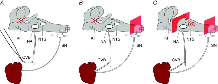
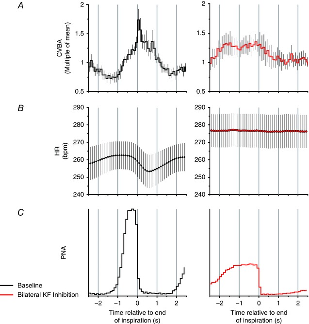
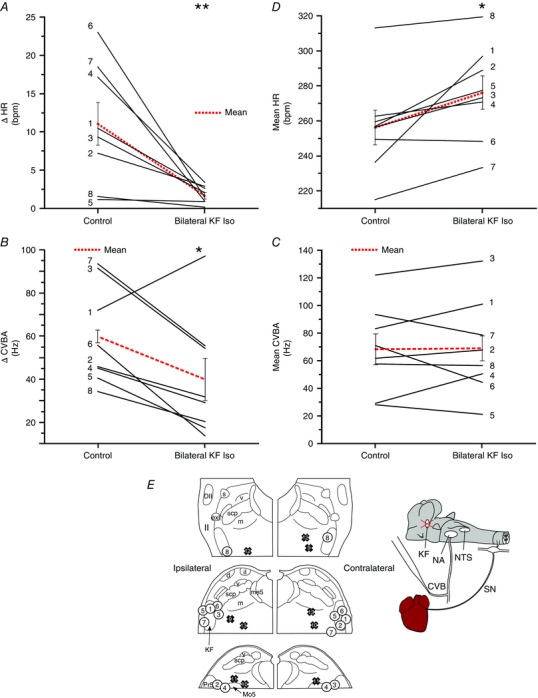
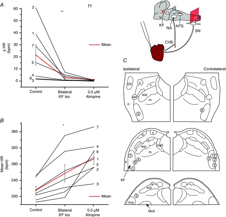
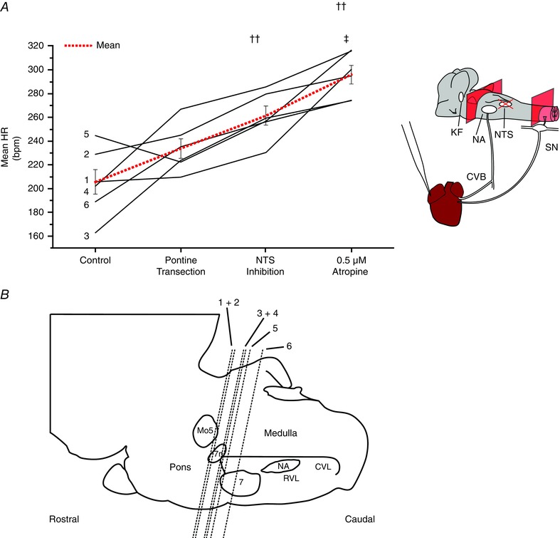
Key points: Cardiac vagal tone is a strong predictor of health, although its central origins are unknown. Respiratory-linked fluctuations in cardiac vagal tone give rise to respiratory sinus arryhthmia (RSA), with maximum tone in the post-inspiratory phase of respiration. In the present study, we investigated whether respiratory modulation of cardiac vagal tone is intrinsically linked to post-inspiratory respiratory control using the unanaesthetized working heart-brainstem preparation of the rat. Abolition of post-inspiration, achieved by inhibition of the pontine Kolliker-Fuse nucleus, removed post-inspiratory peaks in efferent cardiac vagal activity and suppressed RSA, whereas substantial cardiac vagal tone persisted. After transection of the caudal pons, part of the remaining tone was removed by inhibition of nucleus of the solitary tract. We conclude that cardiac vagal tone depends upon at least 3 sites of the pontomedullary brainstem and that a significant proportion arises independently of RSA.

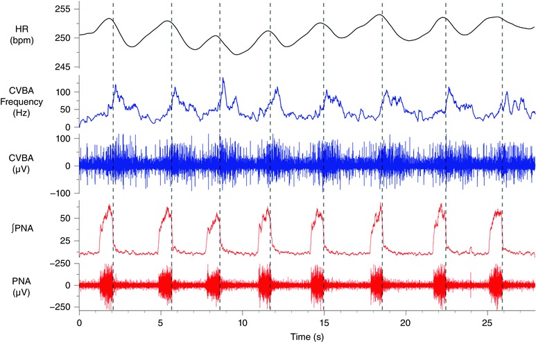
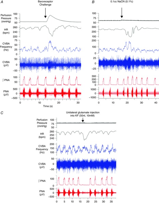
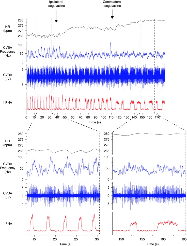
Abstract: Cardiac vagal tone is a strong predictor of health, although its central origins are unknown. The rat working heart-brainstem preparation shows strong cardiac vagal tone and pronounced respiratory sinus arrhythmia. In this preparation, recordings from the cut left cardiac vagal branch showed efferent activity that peaked in post-inspiration, ∼0.5 s before the cyclic minimum in heart rate (HR). We hypothesized that respiratory modulation of cardiac vagal tone and HR is intrinsically linked to the generation of post-inspiration. Neurons in the pontine Kölliker-Fuse nucleus (KF) were inhibited with bilateral microinjections of isoguvacine (50-70 nl, 10 mm) to remove the post-inspiratory phase of respiration. This also abolished the post-inspiratory peak of cardiac vagal discharge (and cyclical HR modulation), although a substantial level of activity remained. In separate preparations with intact cardiac vagal branches but sympathetically denervated by thoracic spinal pithing, cardiac chronotropic vagal tone was quantified by HR compared to its final level after systemic atropine (0.5 μm). Bilateral KF inhibition removed 88% of the cyclical fluctuation in HR but, on average, only 52% of the chronotropic vagal tone. Substantial chronotropic vagal tone also remained after transection of the brainstem through the caudal pons. Subsequent bilateral isoguvacine injections into the nucleus of the solitary tract further reduced vagal tone: remaining sources were untraced. We conclude that cardiac vagal tone depends on neurons in at least three sites of the pontomedullary brainstem, and much of it arises independently of respiratory sinus arrhythmia.


Figures

Figure 1
Figure 1. Variations on the WHBP used to investigate the origins of cardiac vagal tone
Three variations of WHBP were used in the present study. Red crosses indicate sites of microinjections. In the first (A), a cardiac branch of the left thoracic vagus was isolated and severed. This allowed direct, differential recordings of electrical activity of the neurons that project to the heart within this branch. Because making these recordings required that we unilaterally dennervate the heart, these experiments were repeated in preparations with cardiac vagal branches left intact (B). In this case, the thoracic spinal cord was destroyed, removing the confounding chronotopic influence of sympathetic cardiac nerves. Finally (C), the relative contribution of pontine and medullary sources of cardiac vagal tone was assessed by transection of the brainstem at the level of the caudal pons. In each case, the atrial ECG was recorded from the surface of the atrium. The HR was calculated using the interval between adjacent P waves. CVB, cardiac vagal branch; KF, Kölliker‐Fuse; NA, nucleus ambiguus; NTS, nucleus tractus solitarii; SN, cardiac sympathetic nerve. [Colour figure can be viewed at wileyonlinelibrary.com]
Figure 2
Figure 2. Spontaneous phrenic discharge, respiratory‐phasic CVBA and RSA in the rat WHBP
A representative trace from one of twelve animals in which cardiac vagal branch recordings were made. Vertical broken lines mark the rapid decline in PNA (both raw nerve activity and the integrated waveform are depicted) that denotes the onset of post‐inspiration. CVBA (both raw nerve activity and mean frequency of firing are depicted) showed marked respiratory modulation: increasing during the inspiratory period and peaking in the post‐inspiratory period before falling to a minimal level in late expiration. These preparations also displayed a clear RSA. ∫PNA, integrated PNA. [Colour figure can be viewed at wileyonlinelibrary.com]
Figure 3
Figure 3. Reflex activation and KF stimulation: effects on CVBA
Representative traces from three separate experiments. Baroreceptor challenges, produced by a transient increase in the perfusion pressure (A), were associated with marked activation of the cardiac vagal branch and bradycardia. A bolus of sodium cyanide added to the perfusing solution (0.1 ml, 0.1%) evoked an increase in the magnitude and frequency of phrenic discharge and enhanced the firing rate of the cardiac vagal branch during post‐inspiration (B). These post‐inspiratory discharges were associated with periods of bradycardia. Unilateral microinjection of 10 mm glutamate into the KF prolonged the expiratory period (C). CVBA was maintained during the extended expiratory period, producing a bradycardia. ∫PNA, integrated phrenic nerve activity. [Colour figure can be viewed at wileyonlinelibrary.com]
Figure 4
Figure 4. Inhibition of the KF suppresses the post‐inspiratory peak in CVBA and RSA
A representative trace from a single experiment depicting sequential inhibition of the ipsilateral and contralateral KF with isoguvacine (top panel). Times of injection are indicated by labelled arrows. Sections denoted by broken lines depict time‐expanded views of the trace before and after bilateral inhibition of the KF (bottom). Bilateral inhibition of the KF produced an apneustic pattern of discharge in the phrenic nerve. The post‐inspiratory peak in CVBA was abolished along with the RSA. A mild tachycardia was observed. CVBA assumed a tonic pattern of discharge, occasionally interrupted by periods on inhibition in late expiration that were not associated with an obvious change in HR. ∫PNA, integrated PNA. [Colour figure can be viewed at wileyonlinelibrary.com]
Figure 5
Figure 5. Post‐inspiratory cycle‐triggered means of CVBA and HR before and after bilateral inhibition of the KF
Group data: cycle‐triggered means of CVBA (A), HR (B) and PNA (C). Means were triggered by the rapid decline in PNA denoting the onset of post‐inspiration. Mean CVBA at baseline and after inhibition of the KF is displayed as multiples of the mean frequency over the baseline averaging period: this comprised at least 10 respiratory cycles. At baseline (left), the post‐inspiratory peak in CVBA and the nadir in RSA were prominent. After bilateral inhibition of the KF, the post‐inspiratory peak in CVBA was suppressed, mean HR increased and RSA was no longer apparent. Data are the mean ± SEM (minimum of 10 sweeps/experiment; n = 8). [Colour figure can be viewed at wileyonlinelibrary.com]
Figure 6
Figure 6. Inhibition of the KF reduces the magnitude of respiratory fluctuations in cardiac vagal activity and RSA
The magnitude of the RSA (ΔHR) (A) and of cyclical fluctuations in CVBA (ΔCVBA) (B) was reduced by bilateral injection of isoguvacine into the KF. Mean HR (C) showed an increase, whereas mean CVBA (D) was unchanged. Isoguvacine injection sites for each experiment are depicted in (E): numbered circles depict KF injections corresponding to numbered experiments in (A) to (D); numbered crosses depict control injections made 1 mm medial to KF (data not shown; see Results). ΔHR and ΔCVNA were calculated as the difference between the maximum and the minimum values over the course the single respiratory cycle. All parameters were averaged over ≥ 10 cycles/experiment. Data are values from individual experiments (black lines) and are the mean ± SEM (red dashed lines). * P < 0.05, ** P < 0.01, paired t test (n = 8). il, internal lateral parabrachial nucleus; me5, mesencephalic trigeminal tract; Pr5, principal sensory trigeminal nucleus; scp, superior cerebellar peduncle m, medial parabrachial nucleus; s, superior lateral parabrachial nucleus; v, ventral lateral parabrachial nucleus. [Colour figure can be viewed at wileyonlinelibrary.com]
Figure 7
Figure 7. Cardiac vagal tone persists after inhibition of the KF in sympathetically‐disabled preparations
Isoguvacine injections were repeated in seven animals with intact cardiac vagal branches and whose cardiac sympathetic innervation was disabled by destruction of the thoracic spinal cord. Cardiac vagal tone was quantified by comparing the HR before and after isoguvacine injection with its final level after systemic atropine. Inhibition of the KF greatly reduced the magnitude of the RSA (ΔHR) (A). The addition of atropine to the perfusing solution removed any remaining HR fluctuations. On average, inhibition of the KF removed half of chronotropic vagal tone (B). Isoguvacine injection sites for each experiment (numbered circles) (C). Data are values from individual experiments (black lines) and are the mean ± SEM (red dashed lines). ** P < 0.01 compared to control; *** P < 0.001 compared to control, †† P < 0.01 compared to KF isoguvacine pairwise t test (n = 7). il, internal lateral parabrachial nucleus; me5, mesencephalic trigeminal tract; Pr5, principal sensory trigeminal nucleus; scp, superior cerebellar peduncle m, medial parabrachial nucleus; s, superior lateral parabrachial nucleus; v, ventral lateral parabrachial nucleus. [Colour figure can be viewed at wileyonlinelibrary.com]
Figure 8
Figure 8. Pontine and medullary sources of cardiac vagal tone
Cardiac vagal tone was again quantified by comparing the HR before the removal of suspected sources of cardiac vagal tone with its final level after systemic atropine. The magnitude and direction of changes in HR produced by pontine transection varied greatly between experiments, indicating the presence of both excitatory and inhibitory influences on cardiac vagal tone (A). Substantial cardiac vagal tone survived this procedure. Subsequent inhibition of the NTS produced an increase in HR indicating that this is a source of cardiac vagal drive. The level of transection from each experiment can be seen in (B). Data are values from individual experiments (black lines) and are the mean ± SEM (red dashed lines)., ††P < 0.01 compared to pontine transection; ‡P < 0.05 compared to NTS isoguvacine, paired t test (n = 6). 7, facial nucleus; 7n, facial nerve; CVL, caudal ventrolateral medulla; Mo5, motor trigeminal nucleus; RVL, rostral ventrolateral medulla. [Colour figure can be viewed at wileyonlinelibrary.com]

References

    1. Akselrod S, Gordon D, Ubel FA, Shannon DC, Berger AC & Cohen RJ (1981). Power spectrum analysis of heart rate fluctuation: a quantitative probe of beat‐to‐beat cardiovascular control. Science 213, 220–222. - PubMed
    1. Anrep GV, Pascual W & Rossler R (1936. a). respiratory variations of the heart rate. I. – The reflex mechanism of the respiratory arrhythmia. Proc R Soc Lond Ser B – Biol Sci 119, 191–217.
    1. Anrep GV, Pascual W & Rossler R (1936. b). Respiratory variations of the heart rate. II. – The central mechanism of the respiratory arrhythmia and the inter‐relations between the central and the reflex mechanisms. Proc R Soc Lond Ser B – Biol Sci 119, 218–230.
    1. Baekey DM, Dick TE & Paton JFR (2008). Pontomedullary transection attenuates central respiratory modulation of sympathetic discharge, heart rate and the baroreceptor reflex in the in situ rat preparation. Exp Physiol 93, 803–816. - PubMed
    1. Barman SM & Gebber GL (2000). “Rapid” rhythmic discharges of sympathetic nerves: sources, mechanisms of generation, and physiological relevance. J Biol Rhythms 15, 365–379. - PubMed

Publication types

LinkOut - more resources