

Stiffened Extracellular Matrix and Signaling from Stromal Fibroblasts via Osteoprotegerin Regulate Tumor Cell Invasion in a 3-D Tumor in Situ Model
- PMID: 27654881
- PMCID: PMC5264661
- DOI: 10.1007/s12307-016-0188-z
Stiffened Extracellular Matrix and Signaling from Stromal Fibroblasts via Osteoprotegerin Regulate Tumor Cell Invasion in a 3-D Tumor in Situ Model
Abstract
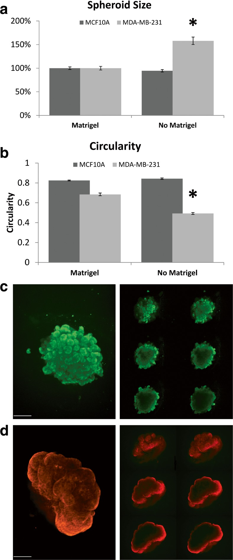
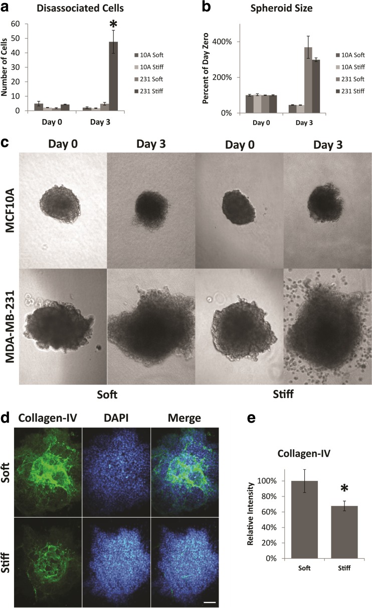
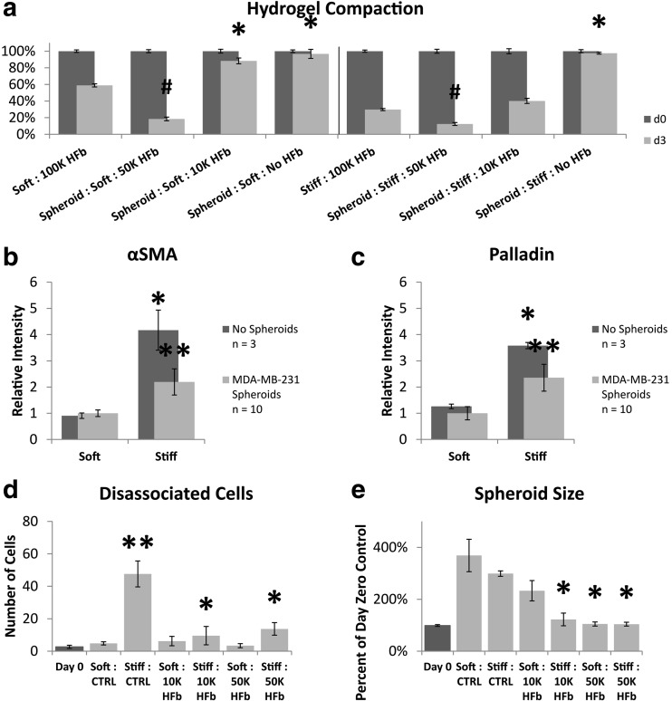
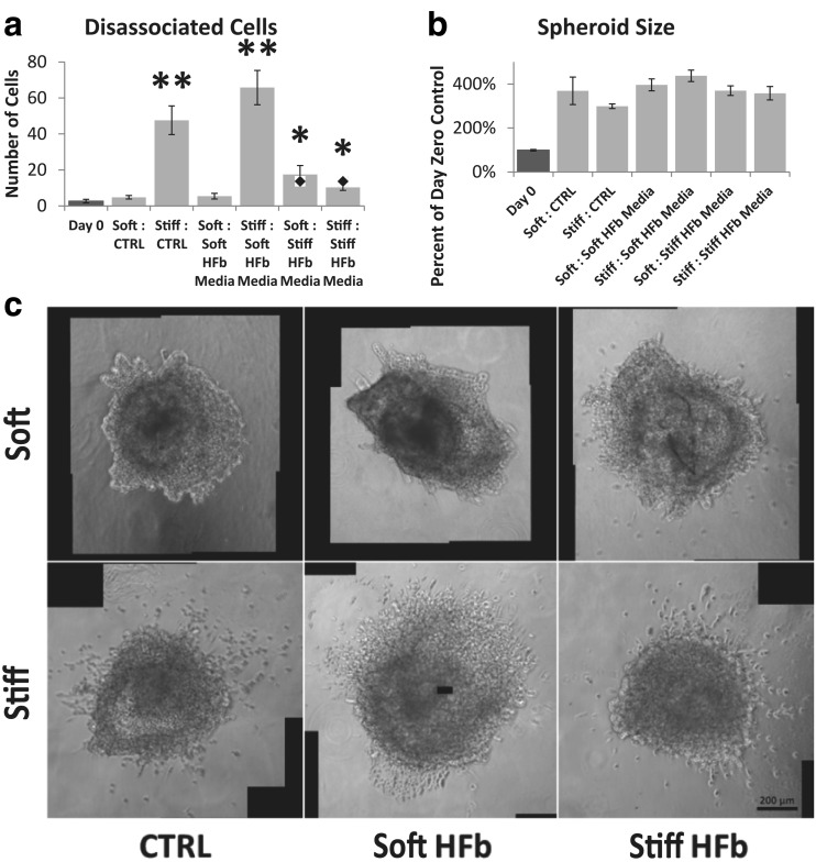
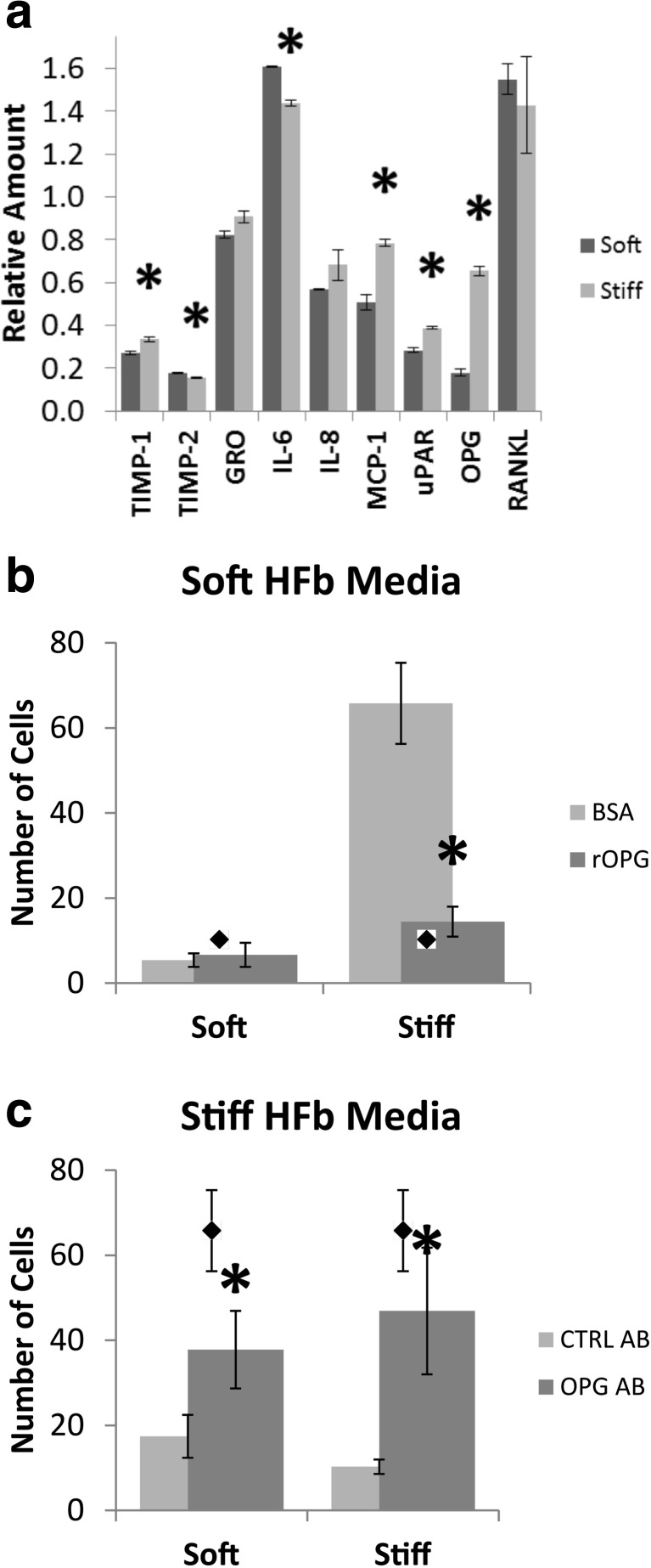
Several changes have been described in the stroma surrounding a tumor, including changes in cellular composition, altered extracellular matrix composition and organization, and increases in stiffness. Tumor cells are influenced by the composition, organization, and mechanical properties of the microenvironment, and by signals from stromal cells. Here we sought to test whether signaling from stromal fibroblasts and/or the small change in stiffness observed in vivo surrounding epithelial tumors regulates tumor cell invasion from a model of a tumor in situ. We generated a novel tumor in situ model system in which a tumor spheroid is encased within a collagen-IV containing membrane and further encased within a collagen-I matrix of in vivo stiffness with or without fibroblasts. Effects of the matrix, fibroblasts or fibroblast signals were determined by observing the invasion of tumor cells into the matrix. Effects of reciprocal tumor cell signaling upon fibroblasts were determined by observing markers of fibroblast activation. We found that a stiffened matrix led to increased dissemination of MDA-MB-231 cells from tumor spheroids when no fibroblasts were present and that MCF10A cells maintained a more normal organization with a stiffened matrix. The presence of fibroblasts, or fibroblast conditioned media, attenuated the effect upon MDA-MB-231 cells. We also observed an attenuation of fibroblast activation associated gene expression in the presence of MDA-MB-231 cells, with a paradoxical increase in activation associated contractile activity. Furthermore, we identified osteoprotegerin as a soluble factor released by fibroblasts in the stiffened environment that is key to the inhibition of cell invasion.
Keywords: 3D hydrogel; Extracellular matrix; Invasion; Mechanotransduction; Tumor in situ.
Conflict of interest statement
Compliance with Ethical Standards Competing Interests The authors declare no competing interests.
Figures
Similar articles
-
Hybrid collagen alginate hydrogel as a platform for 3D tumor spheroid invasion.Acta Biomater. 2018 Jul 15;75:213-225. doi: 10.1016/j.actbio.2018.06.003. Epub 2018 Jun 5. Acta Biomater. 2018. PMID: 29879553 Free PMC article.
-
Mammary fibroblasts remodel fibrillar collagen microstructure in a biomimetic nanocomposite hydrogel.Acta Biomater. 2019 Jan 1;83:221-232. doi: 10.1016/j.actbio.2018.11.010. Epub 2018 Nov 7. Acta Biomater. 2019. PMID: 30414485 Free PMC article.
-
Tumor cell invasion is promoted by interstitial flow-induced matrix priming by stromal fibroblasts.Cancer Res. 2011 Feb 1;71(3):790-800. doi: 10.1158/0008-5472.CAN-10-1513. Epub 2011 Jan 18. Cancer Res. 2011. PMID: 21245098
-
The matrix environmental and cell mechanical properties regulate cell migration and contribute to the invasive phenotype of cancer cells.Rep Prog Phys. 2019 Jun;82(6):064602. doi: 10.1088/1361-6633/ab1628. Epub 2019 Apr 4. Rep Prog Phys. 2019. PMID: 30947151 Review.
-
Tumor-stroma interactions directing phenotype and progression of epithelial skin tumor cells.Differentiation. 2002 Dec;70(9-10):486-97. doi: 10.1046/j.1432-0436.2002.700903.x. Differentiation. 2002. PMID: 12492491 Review.
Cited by
-
The Role of Tumor Microenvironment in Chemoresistance: 3D Extracellular Matrices as Accomplices.Int J Mol Sci. 2018 Sep 20;19(10):2861. doi: 10.3390/ijms19102861. Int J Mol Sci. 2018. PMID: 30241395 Free PMC article.
-
A vascularized breast cancer spheroid platform for the ranked evaluation of tumor microenvironment-targeted drugs by light sheet fluorescence microscopy.Nat Commun. 2024 Apr 27;15(1):3599. doi: 10.1038/s41467-024-48010-z. Nat Commun. 2024. PMID: 38678014 Free PMC article.
-
3D In Vitro Models for Investigating the Role of Stiffness in Cancer Invasion.ACS Biomater Sci Eng. 2023 Jul 10;9(7):3729-3741. doi: 10.1021/acsbiomaterials.0c01530. Epub 2021 Jun 3. ACS Biomater Sci Eng. 2023. PMID: 34081437 Free PMC article. Review.
-
Reshaping in vitro Models of Breast Tissue: Integration of Stromal and Parenchymal Compartments in 3D Printed Hydrogels.Front Bioeng Biotechnol. 2020 Jun 11;8:494. doi: 10.3389/fbioe.2020.00494. eCollection 2020. Front Bioeng Biotechnol. 2020. PMID: 32596217 Free PMC article.
-
The Extracellular Matrix: Its Composition, Function, Remodeling, and Role in Tumorigenesis.Biomimetics (Basel). 2023 Apr 5;8(2):146. doi: 10.3390/biomimetics8020146. Biomimetics (Basel). 2023. PMID: 37092398 Free PMC article. Review.
References
LinkOut - more resources
Full Text Sources
Other Literature Sources
Research Materials
Miscellaneous
