. 2016 Sep 28;283(1839):20161553.
doi: 10.1098/rspb.2016.1553.

Probiotic treatment restores protection against lethal fungal infection lost during amphibian captivity

Affiliations

Probiotic treatment restores protection against lethal fungal infection lost during amphibian captivity

Jordan G Kueneman et al. Proc Biol Sci. .

Abstract

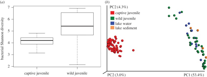
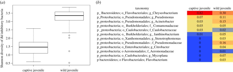
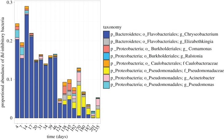
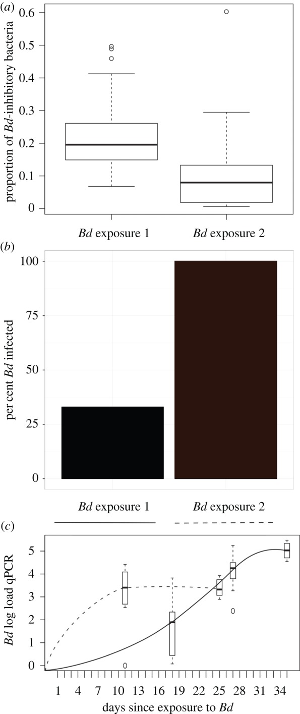
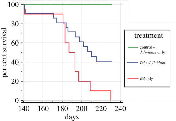
Host-associated microbiomes perform many beneficial functions including resisting pathogens and training the immune system. Here, we show that amphibians developing in captivity lose substantial skin bacterial diversity, primarily due to reduced ongoing input from environmental sources. We combined studies of wild and captive amphibians with a database of over 1 000 strains that allows us to examine antifungal function of the skin microbiome. We tracked skin bacterial communities of 62 endangered boreal toads, Anaxyrus boreas, across 18 time points, four probiotic treatments, and two exposures to the lethal fungal pathogen Batrachochytrium dendrobatidis (Bd) in captivity, and compared these to 33 samples collected from wild populations at the same life stage. As the amphibians in captivity lost the Bd-inhibitory bacteria through time, the proportion of individuals exposed to Bd that became infected rose from 33% to 100% in subsequent exposures. Inoculations of the Bd-inhibitory probiotic Janthinobacterium lividum resulted in a 40% increase in survival during the second Bd challenge, indicating that the effect of microbiome depletion was reversible by restoring Bd-inhibitory bacteria. Taken together, this study highlights the functional role of ongoing environmental inputs of skin-associated bacteria in mitigating a devastating amphibian pathogen, and that long-term captivity decreases this defensive function.

Keywords: Anaxyrus boreas; Batrachochytrium dendrobatidis; amphibian; captivity; microbiome; probiotics.


Figures

Figure 1.
Figure 1.
(a) Shannon diversity of bacterial OTUs on captive and wild juvenile boreal toads. Data shown for captive toads (n = 62) represent the communities sampled on day 4 in captivity and wild toads (n = 30) were sampled in a single high elevation wetland habitat in Colorado. Wild toads had a significantly more diverse skin microbiome compared with captive toads (ANOVA F = 41.47, p < 0.0001). (b) Beta-diversity of captive juveniles toads, wild juveniles, and environmental samples (water and sediment) from the wild. Bacterial communities on captive toads differ dramatically from wild toads and their environment (ANOSIM R2 = 0.8554, p < 0.001). Wild toads were rinsed with sterile water to remove transient environmental microbes [18]. Each point represents the bacterial community, by sample type: red = captive juvenile skin (n = 42), green = wild juvenile skin (n = 42), blue = lake water (n = 13), orange = lake sediment (n = 4). Diversity patterns are visualized using principal coordinate plots of unweighted UniFrac distances.
Figure 2.
Figure 2.
(a) Shannon diversity of Bd-inhibitory taxa found on captive and wild juveniles rarefied to 12 700 sequences per sample. Captive juveniles (n = 62) sampled at day 4 and wild juveniles (n = 30) sampled in the field; ANOVA F = 157.7, p < 0.001. (b) The heatmap depicts the proportional abundance (mean number of sequences per taxon divided by total sequences per host group) of only Bd-inhibitory bacterial OTUs across captive boreal toads and wild boreal toads. Heatmap includes OTUs found with 0.3% or greater of the proportional abundance. Electronic supplementary material, figure S3 includes all inhibitory OTUs found on wild (n = 30 OTUs) and captive (n = 12) juvenile boreal toads. Heatmap is ordered by decreasing taxonomic abundance in captive juvenile individuals. (Online version in colour.)
Figure 3.
Figure 3.
The proportional abundance (as a mean per cent of total sequences per sample) of bacterial taxa on 62 captive boreal toads through time. Bd-inhibitory bacterial taxa are determined as sequence matches to the independent Bd-inhibitory isolates database [10]. Other bacterial taxa that do not match Bd inhibitors are not shown. The proportional abundance of Chryseobacterium decreases through time, ranging from 26% to 0.1% of the total community. (Online version in colour.)
Figure 4.
Figure 4.
The proportion of Bd-inhibitory bacteria on captive toads, the per cent infected, and the pathogen load during the first and second exposures to Bd. (a) The proportion of sequences that match the Bd-inhibitory database found on individuals at the time of Bd exposure 1 (n = 39 toads on day 17) and Bd exposure 2 (n = 31 toads on day 138). There is a higher proportion of the skin community that is Bd inhibitory at the time of first Bd exposure than there was at the time of the second Bd exposure (ANOVA F = 16.22, p < 0.00016). (b) Of 39 toads exposed to Bd in the first trial, 13 (33%) became infected. During the second Bd exposure, 31 out of 31 (100%) toads became infected. (c) Bd load is presented as the number of log-transformed DNA copies (qPCR) through time during Bd exposure 1 (solid line) and Bd exposure 2 (dotted line). Bd load means are calculated from 13 infected individuals in exposure 1 and from 10 infected individuals in exposure 2 (these include the Bd-only infected group and excludes the J. lividum treated group which differed significantly in the second trial, see figure 5; electronic supplementary material, figure S4). (Online version in colour.)
Figure 5.
Figure 5.
Survival of toads treated with Janthinobacterium lividum (J. lividum) prior to exposure to Bd (J. lividum plus Bd, n = 21, blue), toads exposed to Bd only (n = 10, red), toads exposed to J. liv only (n = 10), and controls exposed to a sterile sham treatment (n = 5), combined as green. J. lividum treated toads plus Bd experienced a 40 per cent increase in survival compared to the Bd-only treated toads (Mantel-Cox, χ2 6.021, p = 0.0141). No mortality was observed for control toads or toads treated with J. lividum only. (Online version in colour.)

References

    1. McFall-Ngai M, et al. 2013. Animals in a bacterial world, a new imperative for the life sciences. Proc. Natl Acad. Sci. USA 110, 3229–3236. (10.1073/pnas.1218525110) - DOI - PMC - PubMed
    1. Caballero S, Pamer EG. 2015. Microbiota-mediated inflammation and antimicrobial defense in the intestine. Annu. Rev. Immunol. 33, 227–256. (10.1146/annurev-immunol-032713-120238) - DOI - PMC - PubMed
    1. Blaser M, Bork P, Fraser C, Knight R, Wang J. 2013. The microbiome explored: recent insights and future challenges. Nat. Rev. Microbiol. 11, 213–217. (10.1038/nrmicro2973) - DOI - PubMed
    1. Harris RN, et al. 2009. Skin microbes on frogs prevent morbidity and mortality caused by a lethal skin fungus. ISME J. 3, 818–824. (10.1038/ismej.2009.27) - DOI - PubMed
    1. Lam BA, Walke JB, Vredenburg VT, Harris RN. 2010. Proportion of individuals with anti-Batrachochytrium dendrobatidis skin bacteria is associated with population persistence in the frog Rana muscosa. Biol. Conserv. 143, 529–531. (10.1016/j.biocon.2009.11.015) - DOI