. 2016 Sep 21;36(38):9896-907.
doi: 10.1523/JNEUROSCI.1762-16.2016.

Reduced Efficacy of Anti-Aβ Immunotherapy in a Mouse Model of Amyloid Deposition and Vascular Cognitive Impairment Comorbidity

Affiliations

Reduced Efficacy of Anti-Aβ Immunotherapy in a Mouse Model of Amyloid Deposition and Vascular Cognitive Impairment Comorbidity

Erica M Weekman et al. J Neurosci. .

Abstract

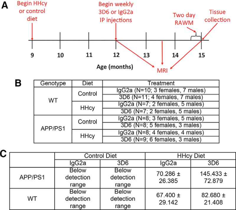
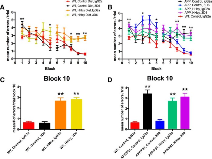
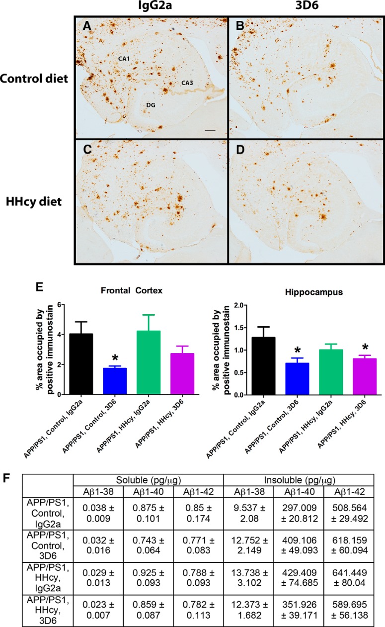
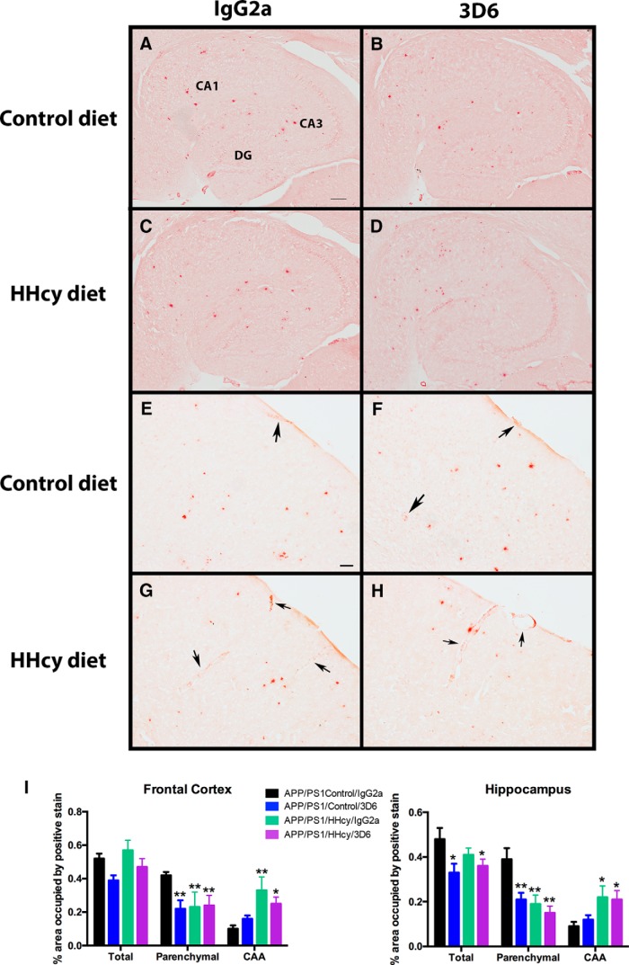
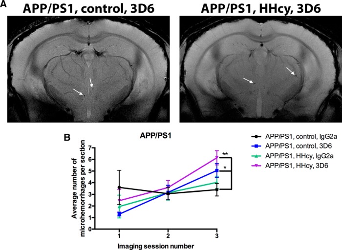
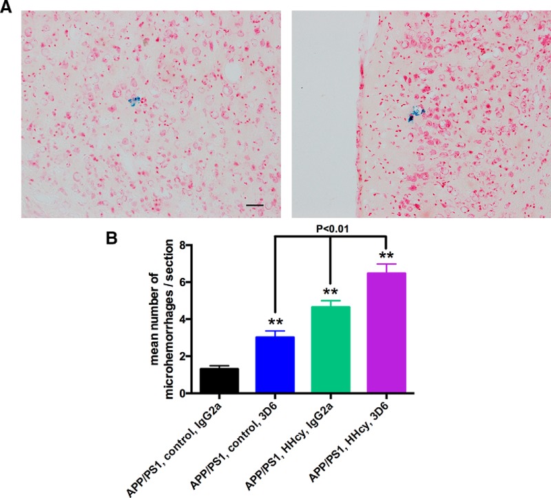
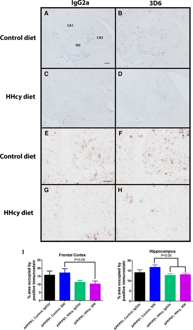
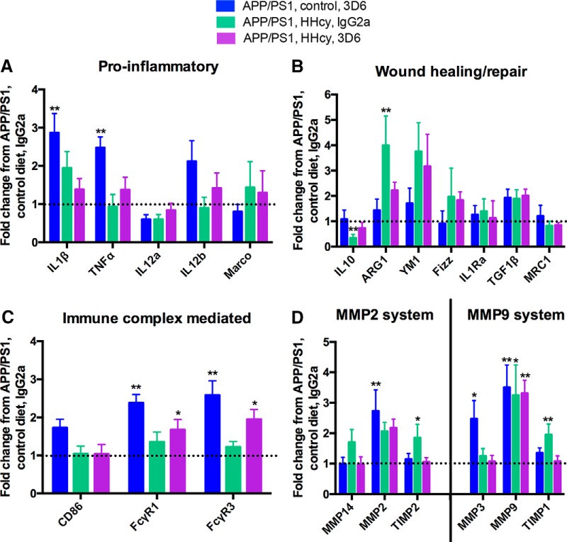
Vascular cognitive impairment and dementia (VCID) is the second most common form of dementia behind Alzheimer's disease (AD). It is estimated that 40% of AD patients also have some form of VCID. One promising therapeutic for AD is anti-Aβ immunotherapy, which uses antibodies against Aβ to clear it from the brain. While successful in clearing Aβ and improving cognition in mice, anti-Aβ immunotherapy failed to reach primary cognitive outcomes in several different clinical trials. We hypothesized that one potential reason the anti-Aβ immunotherapy clinical trials were unsuccessful was due to this high percentage of VCID comorbidity in the AD population. We used our unique model of VCID-amyloid comorbidity to test this hypothesis. We placed 9-month-old wild-type and APP/PS1 mice on either a control diet or a diet that induces hyperhomocysteinemia (HHcy). After being placed on the diet for 3 months, the mice then received intraperotineal injections of either IgG2a control or 3D6 for another 3 months. While we found that treatment of our comorbidity model with 3D6 resulted in decreased total Aβ levels, there was no cognitive benefit of the anti-Aβ immunotherapy in our AD/VCID mice. Further, microhemorrhages were increased by 3D6 in the APP/PS1/control but further increased in an additive fashion when 3D6 was administered to the APP/PS1/HHcy mice. This suggests that the use of anti-Aβ immunotherapy in patients with both AD and VCID would be ineffective on cognitive outcomes.

Significance statement: Despite significant mouse model data demonstrating both pathological and cognitive efficacy of anti-Aβ immunotherapy for the treatment of Alzheimer's disease, clinical trial outcomes have been underwhelming, failing to meet any primary endpoints. We show here that vascular cognitive impairment and dementia (VCID) comorbidity eliminates cognitive efficacy of anti-Aβ immunotherapy, despite amyloid clearance. Further, cerebrovascular adverse events of the anti-Aβ immunotherapy are significantly exacerbated by the VCID comorbidity. These data suggest that VCID comorbidity with Alzheimer's disease may mute the response to anti-Aβ immunotherapy.

Keywords: amyloid; immunotherapy; microglia; microhemorrhage; mixed dementia; vascular cognitive impairment.


Figures

Figure 1.
Figure 1.
Experimental setup. A, Mice were 9 months old when they began either the control or HHcy diet. After being on the diet for 3 months, mice began a 3 month treatment of weekly intraperitoneal injections of 3D6 or IgG2a at 10 mg/kg. MRI was performed 3 months after starting the diet but before intraperitoneal injection treatment, halfway through treatment, and immediately before tissue collection. The 2 d radial arm water maze was also performed before tissue collection. B, Number of animals per experimental group. C, Quantification of plasma total homocysteine levels.
Figure 2.
Figure 2.
The 3D6 treatment does not improve cognition in APP/PS1 mice on the HHcy diet. Two day radial arm water maze data are shown for WT mice (A) and APP/PS1 mice (B). The mean number of errors per trial was calculated for each block; each block is the average of three trials. *p < 0.05, compared with all groups on each graph (one-way ANOVA). **p < 0.01, compared with all groups on each graph (one-way ANOVA). Block 10 data are graphed for the WT mice (C) and the APP/PS1 mice (D). C, **p < 0.01, compared with WT, control, IgG2a and WT, control, and 3D6. D, **p < 0.01, compared with WT, control, IgG2a and APP/PS1, control, and 3D6.
Figure 3.
Figure 3.
Total Aβ is reduced by 3D6 treatment. Representative images of Aβ staining in the hippocampus of APP/PS1, control, and IgG2a (A), APP/PS1, control, and 3D6 (B), APP/PS1, HHcy, and IgG2a (C), and APP/PS1, HHcy, and 3D6 (D). A, The cornu ammonis (CA) 1, CA3, and dentate gyrus (DG) are labeled for orientation. Scale bar: A, 120 μm. E, Quantification of percentage positive stain in the frontal cortex and hippocampus. *p < 0.05, compared with APP/PS1, control, and IgG2a. F, Biochemical quantification of soluble and insoluble Aβ1–38, Aβ1–40, and Aβ1–42 ± SEM.
Figure 4.
Figure 4.
HHcy redistributes amyloid to the vasculature. Representative images of Congo red staining in the hippocampus of APP/PS1, control, and IgG2a (A), APP/PS1, control, and 3D6 (B), APP/PS1, HHcy, and IgG2a (C), and APP/PS1, HHcy, and 3D6 (D). A, The CA1, CA3, and DG are labeled for orientation. Scale bar: A, 200 μm. Representative images of Congo red staining in the cortex of APP/PS1, control, and IgG2a (E), APP/PS1, control, and 3D6 (F), APP/PS1, HHcy, and IgG2a (G), and APP/PS1, HHcy, and 3D6 (H). Scale bar: E, 50 μm. Black arrows indicate Congo staining around a vessel (CAA). I, Quantification of percentage positive stain in the frontal cortex and hippocampus. *p < 0.05, compared with APP/PS1, control, and IgG2a. **p < 0.01, compared with APP/PS1, control, and IgG2a.
Figure 5.
Figure 5.
The 3D6 increases MRI detected microhemorrhages. A, Representative T2* images of 3D6-treated mice. Arrows indicate a microhemorrhage. B, Quantification of the number of microhemorrhages per section over the 3 imaging sessions. *p < 0.05, compared with APP/PS1, control, and IgG2a. **p < 0.01, compared with APP/PS1, control, and IgG2a.
Figure 6.
Figure 6.
The 3D6 and HHcy increase Prussain blue detected microhemorrhages. A, Representative images of Prussian blue-positive microhemorrhages in the frontal cortex. Scale bar, 120 μm. B, Quantification of the mean number of microhemorrhages per section. **p < 0.01, compared with APP/PS1, control, and IgG2a. Black bars represent significant differences between connecting groups.
Figure 7.
Figure 7.
CD11b staining is decreased in the HHcy groups. Representative images of CD11b staining in the hippocampus of APP/PS1, control, and IgG2a (A), APP/PS1, control, and 3D6 (B), APP/PS1, HHcy, and IgG2a (C), and APP/PS1, HHcy, and 3D6 (D). A, The CA1, CA3, and DG are labeled for orientation. Scale bar: A, 120 μm. Higher-magnification images of CD11b staining in the dentate gyrus of APP/PS1, control, and IgG2a (E), APP/PS1, control, and 3D6 (F), APP/PS1, HHcy, and IgG2a (G), and APP/PS1, HHcy, and 3D6 (H). Scale bar: E, 50 μm. I, Quantification of percentage positive stain in the frontal cortex and hippocampus.
Figure 8.
Figure 8.
HHcy reduced inflammatory markers and increased the MMP system markers in both the IgG2a and 3D6 groups. Data are shown as a fold change from APP/PS1, control, and IgG2a. Relative gene expression for proinflammatory markers (A), would healing/repair markers (B), immune complex-mediated markers (C), and MMP2 and MMP9 system markers (D). *p < 0.05, compared with APP/PS1, control, and IgG2a. **p < 0.01, compared with APP/PS1, control, and IgG2a.

References

    1. Alamed J, Wilcock DM, Diamond DM, Gordon MN, Morgan D. Two-day radial-arm water maze learning and memory task: robust resolution of amyloid-related memory deficits in transgenic mice. Nat Protoc. 2006;1:1671–1679. doi: 10.1038/nprot.2006.275. - DOI - PubMed
    1. Alzheimer's Association. 2015 Alzheimer's disease facts and figures. Alzheimers Dement. 2015;11:332–384. doi: 10.1016/j.jalz.2015.02.003. - DOI - PubMed
    1. Bacskai BJ, Kajdasz ST, Christie RH, Carter C, Games D, Seubert P, Schenk D, Hyman BT. Imaging of amyloid-beta deposits in brains of living mice permits direct observation of clearance of plaques with immunotherapy. Nat Med. 2001;7:369–372. doi: 10.1038/85525. - DOI - PubMed
    1. Bard F, Cannon C, Barbour R, Burke RL, Games D, Grajeda H, Guido T, Hu K, Huang J, Johnson-Wood K, Khan K, Kholodenko D, Lee M, Lieberburg I, Motter R, Nguyen M, Soriano F, Vasquez N, Weiss K, Welch B, et al. Peripherally administered antibodies against amyloid beta-peptide enter the central nervous system and reduce pathology in a mouse model of Alzheimer disease. Nat Med. 2000;6:916–919. doi: 10.1038/78682. - DOI - PubMed
    1. Bostom AG, Rosenberg IH, Silbershatz H, Jacques PF, Selhub J, D'Agostino RB, Wilson PW, Wolf PA. Nonfasting plasma total homocysteine levels and stroke incidence in elderly persons: the Framingham Study. Ann Intern Med. 1999;131:352–355. doi: 10.7326/0003-4819-131-5-199909070-00006. - DOI - PubMed

Publication types

MeSH terms

LinkOut - more resources