Review
. 2016 Sep 5:10:71.
doi: 10.3389/fncir.2016.00071. eCollection 2016.

The Wiring of Developing Sensory Circuits-From Patterned Spontaneous Activity to Synaptic Plasticity Mechanisms

Affiliations
Review

The Wiring of Developing Sensory Circuits-From Patterned Spontaneous Activity to Synaptic Plasticity Mechanisms

Alexandra H Leighton et al. Front Neural Circuits. .

Abstract

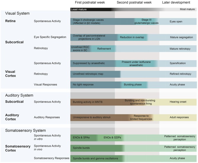
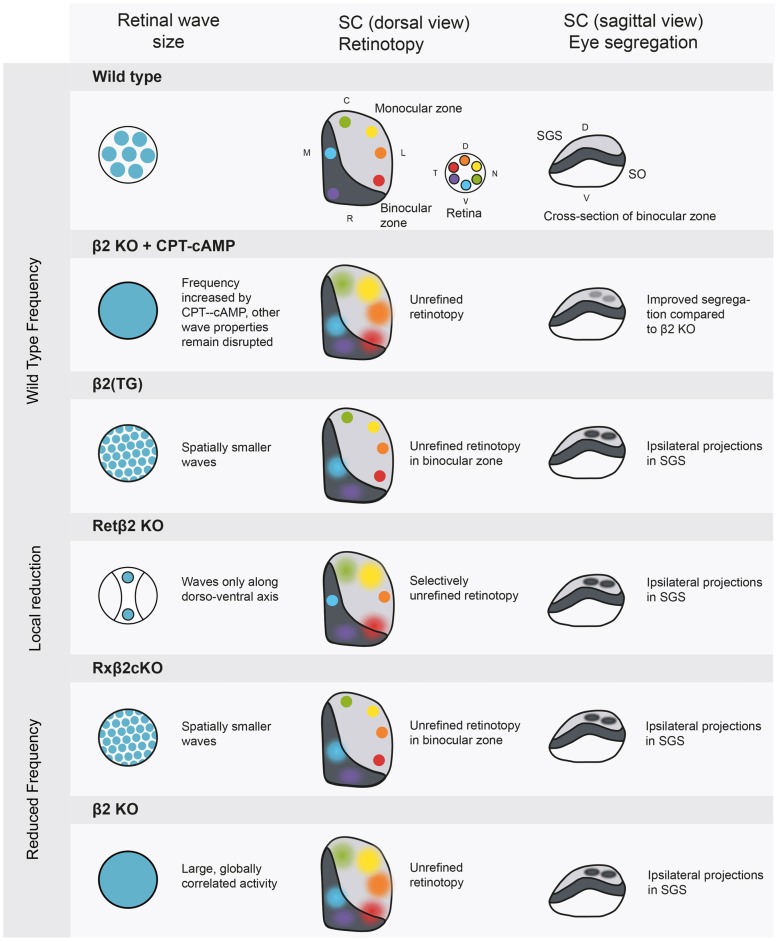
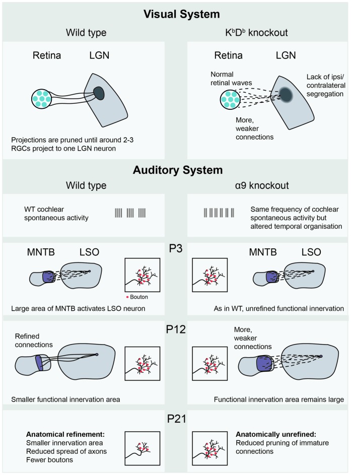
In order to accurately process incoming sensory stimuli, neurons must be organized into functional networks, with both genetic and environmental factors influencing the precise arrangement of connections between cells. Teasing apart the relative contributions of molecular guidance cues, spontaneous activity and visual experience during this maturation is on-going. During development of the sensory system, the first, rough organization of connections is created by molecular factors. These connections are then modulated by the intrinsically generated activity of neurons, even before the senses have become operational. Spontaneous waves of depolarizations sweep across the nervous system, placing them in a prime position to strengthen correct connections and weaken others, shaping synapses into a useful network. A large body of work now support the idea that, rather than being a mere side-effect of the system, spontaneous activity actually contains information which readies the nervous system so that, as soon as the senses become active, sensory information can be utilized by the animal. An example is the neonatal mouse. As soon as the eyelids first open, neurons in the cortex respond to visual information without the animal having previously encountered structured sensory input (Cang et al., 2005b; Rochefort et al., 2011; Zhang et al., 2012; Ko et al., 2013). In vivo imaging techniques have advanced considerably, allowing observation of the natural activity in the brain of living animals down to the level of the individual synapse. New (opto)genetic methods make it possible to subtly modulate the spatio-temporal properties of activity, aiding our understanding of how these characteristics relate to the function of spontaneous activity. Such experiments have had a huge impact on our knowledge by permitting direct testing of ideas about the plasticity mechanisms at play in the intact system, opening up a provocative range of fresh questions. Here, we intend to outline the most recent descriptions of spontaneous activity patterns in rodent developing sensory areas, as well as the inferences we can make about the information content of those activity patterns and ideas about the plasticity rules that allow this activity to shape the young brain.

Keywords: auditory system development; developmental biology; plasticity mechanisms; spontaneous activity; synaptic plasticity; visual system development.


Figures

Figure 1
Figure 1
Spontaneous and evoked activity during early postnatal development in mice and rats, and the changes in patterning that occur during this time. Visual system: (McLaughlin et al., ; Cang et al., ; Firth et al., ; Torborg and Feller, ; Demas et al., ; Rochefort et al., ; Colonnese et al., ; Siegel et al., 2012). Auditory system (Geal-Dor et al., ; Kandler and Gillespie, ; Sonntag et al., ; Tritsch et al., ; Froemke and Jones, 2011) Somatosensory system (Minlebaev et al., ; Allène et al., ; Allene and Cossart, ; Colonnese et al., 2010). ENO, early network oscillations; SPA, synchronized plateau assemblies; GDPs, giant depolarizing potentials; RGC, retinal ganglion cell; LGN, lateral geniculate nucleus; MNTB, medial nucleus of the trapezoid body; SC, superior colliculus.
Figure 2
Figure 2
Manipulations of spontaneous activity frequency and wave size and the consequences for retinotopy and eye-specific segregation in the SC. The SC has a binocular and a monocular region, and contains a retinotopic map in a mirror image of the retina. The most superficial layer of the SC is the stratum griseum superficial (SGS), which is targeted only by axons from the contralateral eye. The stratum opticum (SO) contains ipsilateral projections in wild type animals. Wild type: retinal activity in the wild type mouse. β2 knockout+ cAMP-CPT: this manipulation increases frequency to wild type levels (Burbridge et al., 2014), rescuing eye specific segregation but not retinotopy. β2 (TG): truncated waves as in the β2 (TG) mouse disturb eye-specific segregation (Xu et al., 2011). Retβ2-KO: partially disrupting wave activity also has spatially selective consequences for retinotopy (Burbridge et al., 2014). Rxβ2-KO: reducing wave frequency and size disturbs segregation (Xu et al., 2015) and retinotopy in the binocular zone. β2 KO: the whole body β2 knockout has low frequency activity over large areas of the retina, leading to unrefined retinotopy and disrupted eye specific segregation. SGS, stratum griseum superficiale; SO, stratum opticum; D, dorsal; V, ventral; C, caudal; R, rostral; T, temporal; N, nasal.
Figure 3
Figure 3
Recent empirical evidence for Hebbian and homeostatic plasticity mechanisms mediated by spontaneous activity. The KbDb−/− knockout (Lee et al., 2014) shows lack of LTD and altered retinogeniculate projections, in which many weak projections connect the retina to the LGN. In the auditory system, the MNTB-LSO projection in the α9 subunit knockout (Clause et al., 2014) shows both functional and anatomical consequences of altered cochlear spontaneous activity, occurring at different postnatal ages. LGN, Lateral geniculate nucleus; RGC, retinal ganglion cell; WT, wild-type; MNTB, medial nucleus of the trapezoid body; LSO, lateral superior olive; P3, postnatal day 3.

References

    1. Ackman J. B., Burbridge T. J., Crair M. C. (2012). Retinal waves coordinate patterned activity throughout the developing visual system. Nature 490, 219–225. 10.1038/nature11529 - DOI - PMC - PubMed
    1. Ackman J. B., Crair M. C. (2014). Role of emergent neural activity in visual map development. Curr. Opin. Neurobiol. 24, 166–175. 10.1016/j.conb.2013.11.011 - DOI - PMC - PubMed
    1. Adelsberger H., Garaschuk O., Konnerth A. (2005). Cortical calcium waves in resting newborn mice. Nat. Neurosci. 8, 988–990. 10.1038/nn1502 - DOI - PubMed
    1. Allène C., Cattani A., Ackman J. B., Bonifazi P., Aniksztejn L., Ben-Ari Y., et al. . (2008). Sequential generation of two distinct synapse-driven network patterns in developing neocortex. J. Neurosci. 28, 12851–12863. 10.1523/jneurosci.3733-08.2008 - DOI - PMC - PubMed
    1. Allene C., Cossart R. (2010). Early NMDA receptor-driven waves of activity in the developing neocortex: physiological or pathological network oscillations? J. Physiol. 588, 83–91. 10.1113/jphysiol.2009.178798 - DOI - PMC - PubMed