Review
. 2016 Sep 6:7:342.
doi: 10.3389/fimmu.2016.00342. eCollection 2016.

Development and Function of Secondary and Tertiary Lymphoid Organs in the Small Intestine and the Colon

Affiliations
Review

Development and Function of Secondary and Tertiary Lymphoid Organs in the Small Intestine and the Colon

Manuela Buettner et al. Front Immunol. .

Abstract

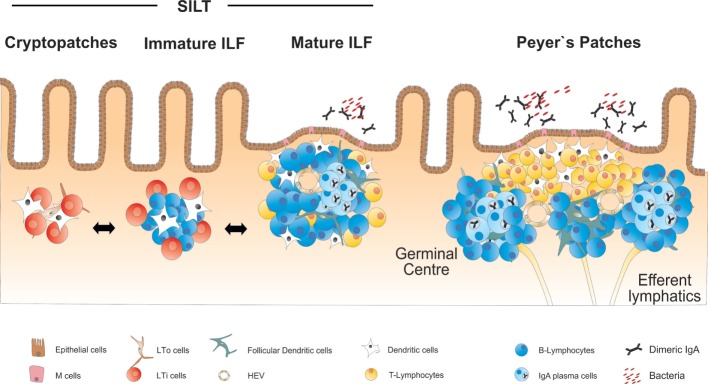
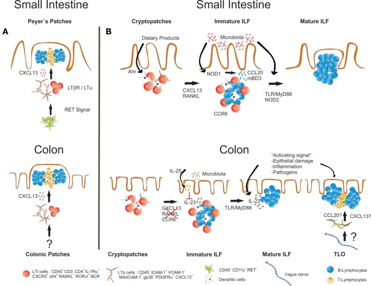
The immune system of the gut has evolved a number of specific lymphoid structures that contribute to homeostasis in the face of microbial colonization and food-derived antigenic challenge. These lymphoid organs encompass Peyer's patches (PP) in the small intestine and their colonic counterparts that develop in a programed fashion before birth. In addition, the gut harbors a network of lymphoid tissues that is commonly designated as solitary intestinal lymphoid tissues (SILT). In contrast to PP, SILT develop strictly after birth and consist of a dynamic continuum of structures ranging from small cryptopatches (CP) to large, mature isolated lymphoid follicles (ILF). Although the development of PP and SILT follow similar principles, such as an early clustering of lymphoid tissue inducer (LTi) cells and the requirement for lymphotoxin beta (LTβ) receptor-mediated signaling, the formation of CP and their further maturation into ILF is associated with additional intrinsic and environmental signals. Moreover, recent data also indicate that specific differences exist in the regulation of ILF formation between the small intestine and the colon. Importantly, intestinal inflammation in both mice and humans is associated with a strong expansion of the lymphoid network in the gut. Recent experiments in mice suggest that these structures, although they resemble large, mature ILF in appearance, may represent de novo-induced tertiary lymphoid organs (TLO). While, so far, it is not clear whether intestinal TLO contribute to the exacerbation of inflammatory pathology, it has been shown that ILF provide the critical microenvironment necessary for the induction of an effective host response upon infection with enteric bacterial pathogens. Regarding the importance of ILF for intestinal immunity, interfering with the development and maturation of these lymphoid tissues may offer novel means for manipulating the immune response during intestinal infection or inflammation.

Keywords: cryptopatch; isolated lymphoid follicles; large intestine; lymphoid tissue inducer cells; small intestine; tertiary lymphoid organs.


Figures

Figure 1
Figure 1
Overview on the anatomy and structure of CP, ILF, and PP in the small intestine. SILT consists of a dynamic continuum of structures ranging from small cryptopatches (CP) to large mature isolated lymphoid follicles (ILF). CP start to develop into immature ILF by recruiting B cells. Mature ILF contain one big B cell follicle and develop germinal centers, vascular structures, and a follicle-associated epithelium. PP represent the most structured lymphoid organs in the intestine, containing several B cell follicles and distinct T and B cell areas.
Figure 2
Figure 2
Similarities and differences in the development of small intestinal and colonic lymphoid tissues. (A) Development of both PP and colonic patches starts before birth with the clustering of LTα1β2-expressing IL-7Rα+RORγt+ LTi cells together with LTo cells. LTi cell clustering and further development of both PP and colonic patches requires expression of CXCL13 and is dependent on LT-signaling. Whether the initial clustering of LTi cells in the colon depends on RET-expressing IL7RαCD11c+ cells, as it has been described for PP development, is not known. (B) ILF formation in both small intestine and colon starts after birth with the clustering of IL-7Rα+RORγt+ LTi cells in CP. Maintenance and further development of these structures into ILF requires LT signaling in both small intestine and the colon. In the small intestine, also signaling via the Ahr and the expression of CXCL13, RANKL and CCR6 is required, and the commensal microbiota induce ILF formation and maturation via activation of signaling pathways that include NOD1, NOD2, TLRs, and MyD88. In the colon, ILF formation does not seem to critically depend on CXCL13, RANKL, and CCR6. There, the presence of commensal microbiota rather inhibits ILF maturation, probably by inducing IL-25 production from the epithelium, which in turn diminishes the secretion of IL-23, a cytokine that specifically promotes ILF maturation in the colon. Signals associated with intestinal inflammation or infection with enteric pathogens also induce ILF formation and maturation. Intestinal inflammation or infection may also result in the induction of TLO that form independently of LTi cells. TLO may be discriminated from ILF by structural differences such as the presence of T cell areas. A role for the intestinal nervous system for TLO induction in intestinal inflammation has been suggested.

References

    1. Chikly B. Who discovered the lymphatic system. Lymphology (1997) 30:186–93. - PubMed
    1. Boehm T, Bleul CC. The evolutionary history of lymphoid organs. Nat Immunol (2007) 8:131–5.10.1038/ni1435 - DOI - PubMed
    1. Miller JF. Immunological function of the thymus. Lancet (1961) 2:748–9.10.1016/S0140-6736(61)90693-6 - DOI - PubMed
    1. Pitzalis C, Jones GW, Bombardieri M, Jones SA. Ectopic lymphoid-like structures in infection, cancer and autoimmunity. Nat Rev Immunol (2014) 14:447–62.10.1038/nri3700 - DOI - PubMed
    1. van de Pavert SA, Olivier BJ, Goverse G, Vondenhoff MF, Greuter M, Beke P, et al. Chemokine CXCL13 is essential for lymph node initiation and is induced by retinoic acid and neuronal stimulation. Nat Immunol (2009) 10:1193–9.10.1038/ni.1789 - DOI - PMC - PubMed