. 2016:2016:9130754.
doi: 10.1155/2016/9130754. Epub 2016 Aug 31.

Cystic and Papillary Neoplasm at the Hepatic Hilum Possibly Originating in the Peribiliary Glands

Affiliations

Cystic and Papillary Neoplasm at the Hepatic Hilum Possibly Originating in the Peribiliary Glands

Takashi Miyata et al. Case Rep Pathol. 2016.

Abstract

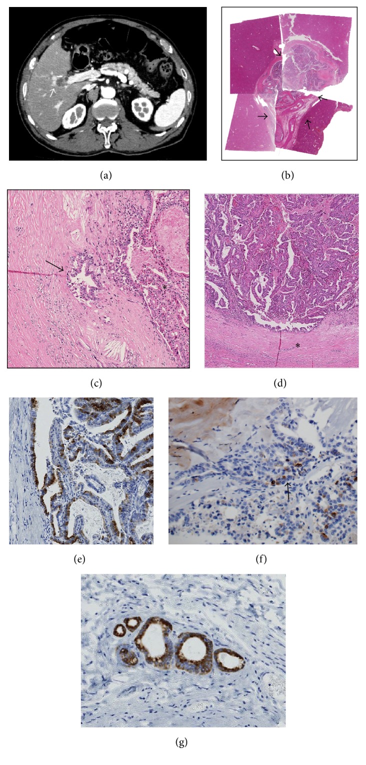
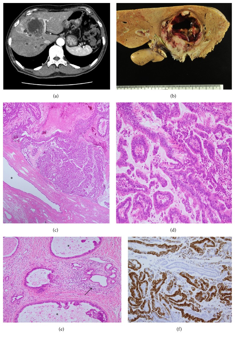
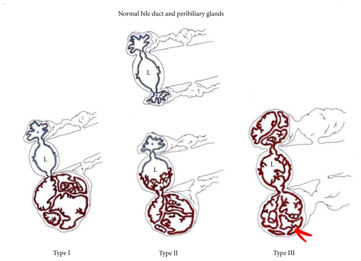
Cystic neoplasms of the liver are divided into two types: mucinous cystic neoplasm and cystic intraductal papillary neoplasm of the bile duct. We herein report two cases of cystic and papillary neoplasm of the liver which differed from the abovementioned types. Case 1. A 70-year-old man. Radiologically, a cystic tumor measuring 20 mm in diameter was found at the hepatic hilum. Right hepatectomy was performed under a diagnosis of intrahepatic cholangiocarcinoma (iCCA) based on the imaging findings. Case 2. A 70-year-old man. Radiologically, a cystic tumor measuring 60 mm in diameter was found at the hepatic hilum. Under a diagnosis of iCCA, left hepatic trisectionectomy was performed. In both cases, endoscopic retrograde cholangiography did not demonstrate communication between the cystic tumor and adjacent bile ducts. Pathologically, these two tumors were cystic neoplasms located at the hepatic hilum and were morphologically characterized by an intracystic papillary neoplasm composed of diffuse high-grade dysplasia and associated with an invasive carcinoma. Ovarian-like stroma was not found in the capsule of these tumors. Interestingly, there were peribiliary glands near these tumors, and MUC6 was expressed in these papillary neoplasms as well as in the peribiliary glands. These neoplasms might have arisen from the peribiliary glands.


Figures

Figure 1
Figure 1
Case  1. (a) Contrast-enhanced CT shows a cystic lesion (arrow) (20 mm in diameter) at the confluence of the anterior and right bile duct with ring enhancement. There is no dilatation of the intrahepatic bile duct, except for slight dilatation of the proximal branches of the anterior branch of the bile duct. (b) The tumor (20 × 17 mm) is cystic and is filled with neoplastic cells and surrounded by a fibrous cyst wall (arrows). Loupe picture of the tumor (H&E staining). (c) Focal invasion of carcinoma is found in the fibrous capsule (arrow). Original magnification, 100x; H&E staining. (d) A papillary neoplasm composed of high-grade dysplasia with fine fibrovascular cores fills the cystic space of the tumor.   Fibrous capsule. Original magnification, 30x; H&E staining. (e) The papillary neoplasm is positive for MUC6. Original magnification, 100x; immunostaining for MUC6. (f) The invasive carcinoma in the fibrous capsule is positive for MUC6 (arrow). Original magnification, 100x; immunostaining for MUC6. (g) The peribiliary glands (nonneoplastic) adjacent to the tumor are also positive for MUC6. Original magnification, 100x; immunostaining for MUC6.
Figure 2
Figure 2
Case  2. (a) Contrast-enhanced CT shows a cystic lesion (60 mm in diameter) with an enhanced wall near the hepatic hilus. (b) A cystic tumor with fibrous wall is found in segment 4, adjacent to the hepatic hilus. (c) Histologically, well-differentiated papillary adenocarcinoma with thin fibrovascular cores fills the cystic space of the tumor.  Surrounding nonneoplastic bile duct. Original magnification, 30x; H&E staining. (d) Papillary carcinoma with columnar epithelia and fine fibrovascular cores. Original magnification, 120x; H&E staining. (e) Invasion of carcinoma () in the periductal tissue of adjacent bile ducts. The arrow denotes the nonneoplastic peribiliary glands. Original magnification, 100x; H&E staining. (f) Papillary carcinoma is positive for MUC6. Original magnification, 100x; immunostaining for MUC6.
Figure 3
Figure 3
A schematic diagram of cystic and papillary neoplasms involving the peribiliary glands (PGs). The blue epithelium indicates nonneoplastic glands, and brown epithelium indicates neoplastic glands. Red indicates an invasion of carcinoma arising in the neoplasm of the PGs. L: bile duct lumen;  unaffected PGs. Type I shows neoplastic involvement confined to the PG, and type II shows main involvement of the PGs with partial secondary involvement of the adjacent bile duct. Type III reflects extensive involvement of the PGs with simultaneous involvement of the bile ducts, which may appear grossly as peribiliary cysts in the perihilar regions.

References

    1. Nakanuma Y., Hoso M., Sanzen T., Sasaki M. Microstructure and development of the normal and pathologic biliary tract in humans, including blood supply. Microscopy Research and Technique. 1997;38(6):552–570. doi: 10.1002/(sici)1097-0029(19970915)38:6<552::aid-jemt2>3.0.co;2-h. - DOI - PubMed
    1. Nakanuma Y., Curabo M. O., Franceschi S., et al. WHO Classification of Tumours of the Digestive System World Health Organization Classification of Tumours. 4th. chapter 10. Lyon, France: IARC Press; 2010. Intrahepatic cholangiocarcinoma; pp. 217–224.
    1. Tsui W. M. S., Adsay N. V., Crawford J. M., Hruban R., Kloppel G., Wee A. Mucinous cystic neoplasms of the liver. In: Bosman F. T., Carneiro F., Hruban R. H., Theise N. D., editors. World Health Organization Classification of Tumours. Pathology and Genetics of Tumours of the Digestive System. 4th. chapter 10. Lyon, France: IARC Press; 2010. pp. 236–238.
    1. Chen T. C., Nakanuma Y., Zen Y., et al. Intraductal papillary neoplasia of the liver associated with hepatolithiasis. Hepatology. 2001;44(5):1333–1343. - PubMed
    1. Nakanuma Y., Kakuda Y. Pathologic classification of cholangiocarcinoma: new concepts. Best Practice and Research: Clinical Gastroenterology. 2015;29(2):277–293. doi: 10.1016/j.bpg.2015.02.006. - DOI - PubMed

LinkOut - more resources