. 2016 May 23;5(3):e507-11.
doi: 10.1016/j.eats.2016.02.012. eCollection 2016 Jun.

A Fluoroscopy-Free Technique for Percutaneous Screw Positioning During Arthroscopic Treatment of Depression Tibial Plateau Fractures

Affiliations

A Fluoroscopy-Free Technique for Percutaneous Screw Positioning During Arthroscopic Treatment of Depression Tibial Plateau Fractures

Mathieu Thaunat et al. Arthrosc Tech. .

Abstract

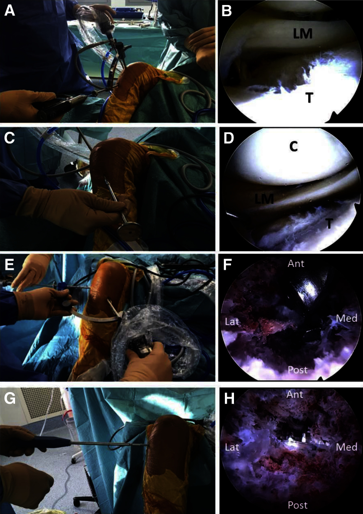
This article aims to describe a simple and reliable technique that helps in positioning the cannulated percutaneous screws during fixation of depression-type tibial plateau fractures. After fracture reduction under arthroscopic control, an outside-in anterior cruciate ligament femoral guide is introduced through the tibial cortical metaphyseal window and positioned under endoscopic control just underneath the elevated fragment. When proper height is achieved, a guide pin is drilled from lateral to medial through the sleeve, 1 to 2 cm distal to the articular surface of the depressed fragment. The cannulated screw can then be introduced under endoscopic control, without fluoroscopic assistance, just under the previously elevated joint surface. This technique ensures optimal placement of the cannulated screw in the middle of the bony tunnel to obtain optimal subchondral bone support during fixation of the depressed tibial plateau fracture.


Figures

Fig 1
Fig 1
Our simple trick allows the surgeon to avoid the use of fluoroscopy intraoperatively to position the cannulated screw for fixation of a depression lateral tibial plateau fracture. After reduction of the fragment, an outside-in anterior cruciate ligament femoral guide, set at maximum amplitude with a 115° to 120° aiming device, is introduced through the tibial cortical metaphyseal window and positioned under endoscopic control just underneath the elevated fragment. This ensures optimal placement of the screw in the middle of the bony tunnel just underneath the elevated fragment to obtain an optimal rafter effect.
Fig 2
Fig 2
Surgical technique. (A, B) The patient is placed in the supine position on a standard table. A tibia-specific jig (Tibial Plateau Fracture Management System) is positioned arthroscopically, through the anteromedial portal. When satisfactory orientation is achieved, a guide pin is drilled toward the deepest point of the fracture. (C, D) A cannulated tamp (Arthrex) is introduced through the tibial cortical window, and under arthroscopic guidance, the depressed fragment is carefully elevated until satisfactory reduction is achieved. (E, F) An outside-in anterior cruciate ligament femoral guide (Arthrex), set at maximum amplitude with a 115° to 120° aiming device, is introduced through the tibial cortical metaphyseal window and positioned under endoscopic control just underneath the elevated fragment. (G, H) A 7-mm cannulated cancellous screw with an 16- or 32-mm thread length is then introduced over the guide pin, after drilling and tapping (Magna-FX cannulated screw system), under endoscopic control. (Ant, anterior; C, condyle; Lat, lateral; LM, lateral meniscus; Med, medial; Post, posterior; T, tibia.)
Fig 3
Fig 3
Preoperative and postoperative radiographs of 2 cases of Schatzker type 3 depressed lateral tibial plateau fracture. (A, C) Tibial plateau fractures (arrows) occasionally are difficult to appreciate with standard radiographs. The preferred examination consists of radiographs in multiple obliquities of the knee. (B, D) Postoperative anteroposterior views after 7-mm cannulated screws (Magna-FX cannulated screw system) were used as a rafter to support the articular surface.

Similar articles

Cited by

References

    1. Caspari R.B., Hutton P.M., Whipple T.L., Meyers J.F. The role of arthroscopy in the management of tibial plateau fractures. Arthroscopy. 1985;1:76–82. - PubMed
    1. Jennings J.E. Arthroscopic management of tibial plateau fractures. Arthroscopy. 1985;1:160–168. - PubMed
    1. Burdin G. Arthroscopic management of tibial plateau fractures: Surgical technique. Orthop Traumatol Surg Res. 2013;99(suppl):S208–S218. - PubMed
    1. Suganuma J., Akutsu S. Arthroscopically assisted treatment of tibial plateau fractures. Arthroscopy. 2004;20:1084–1089. - PubMed
    1. Lubowitz J.H., Elson W.S., Guttmann D. Part I: Arthroscopic management of tibial plateau fractures. Arthroscopy. 2004;20:1063–1070. - PubMed

LinkOut - more resources