. 2016 Jun 30;5(8):626-634.
doi: 10.1016/j.molmet.2016.06.013. eCollection 2016 Aug.

Lipolysis sensation by white fat afferent nerves triggers brown fat thermogenesis

Affiliations

Lipolysis sensation by white fat afferent nerves triggers brown fat thermogenesis

John T Garretson et al. Mol Metab. .

Abstract

Objective: Metabolic challenges, such as a cold environment, stimulate sympathetic neural efferent activity to white adipose tissue (WAT) to drive lipolysis, thereby increasing the availability of free fatty acids as one source of fuel for brown adipose tissue (BAT) thermogenesis. WAT is also innervated by sensory nerve fibers that network to metabolic brain areas; moreover, activation of these afferents is reported to increase sympathetic nervous system outflow. However, the endogenous stimuli sufficient to drive WAT afferents during metabolic challenges as well as their functional relation to BAT thermogenesis remain unknown.

Method: We tested if local WAT lipolysis directly activates WAT afferent nerves, and then assessed whether this WAT sensory signal affected BAT thermogenesis in Siberian hamsters (Phodopus sungorus).

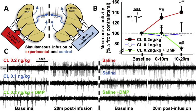
Results: 2-deoxyglucose, a sympathetic nervous system stimulant, caused β-adrenergic receptor dependent increases in inguinal WAT (IWAT) afferent neurophysiological activity. In addition, direct IWAT injections of the β3-AR agonist CL316,243 dose-dependently increased: 1) phosphorylation of IWAT hormone sensitive lipase, an indicator of SNS-stimulated lipolysis, 2) expression of the neuronal activation marker c-Fos in dorsal root ganglion neurons receiving sensory input from IWAT, and 3) IWAT afferent neurophysiological activity, an increase blocked by antilipolytic agent 3,5-dimethylpyrazole. Finally, we demonstrated that IWAT afferent activation by lipolysis triggers interscapular BAT thermogenesis through a neural link between these two tissues.

Conclusions: These data suggest IWAT lipolysis activates local IWAT afferents triggering a neural circuit from WAT to BAT that acutely induces BAT thermogenesis.

Keywords: Adipose innervation; BAT thermogenesis; Denervation; Lipolysis; WAT sensory.


Figures

Figure 1
Figure 1
Glucoprivation increases IWAT multiunit nerve spiking in a β-AR dependent fashion. 2DG (500 mg/kg, i.p.) increased IWAT multiunit spiking, an effect blocked by pretreatment with β-AR antagonist propranolol. Bars represent number of spikes per 30 s of IWAT afferents in hamsters that received either propranolol (40 mg/kg, s.c.) or saline vehicle (s.c.) pretreatment prior to 2DG application. * different from saline pretreatment, p < 0.05.
Figure 2
Figure 2
Intra-adipose β3-AR agonism increases IWAT multiunit nerve activity. (A) Diagrammatic representation of real-time in vivo bilateral multiunit electrophysiology of WAT afferent nerves. Afferent nerves resected from ventrally exposed IWAT (blue curved lines), cut proximal to electrodes (gray hook pairs) to eliminate efferent interference, and analyzed for multiunit spike frequency over time. Symmetrically placed infusion needles indicate bilateral simultaneous infusion of lipolytic drug CL316,243 (CL, β3-AR agonist) and its sterile saline vehicle (no drug) in the contralateral negative control pad. (B) CL dose-dependently increased IWAT multiunit spiking, an effect blocked by pretreatment with the antilipolytic drug 3,5-dimethylpyrazole (DMP, 12 mg/kg, i.p). Data are expressed as a percentage change of activity from CL-infused pad over contralateral to eliminate whole animal IWAT nerve variability caused by the recording/injection procedure. Inset is a representative single spike to display acquired waveforms. (C) Representative (20 s) traces from IWAT afferents and contralateral counterparts at baseline and 20 min post-infusion where the ipsilateral IWAT was infused with CL at either 0.2 ng/kg (top), 0.1 ng/kg (middle), or 0.2 ng/kg 20 min after pretreatment with antilipolytic drug DMP (bottom). CL injections were counterbalanced by side to control for any possible side bias. Scale bars on first trace apply to all traces. Dotted line separates ipsilateral vs contralateral IWAT afferent recordings from each of the three representative animals. * different from contralateral control; # different from baseline activity, p < 0.05.
Figure 3
Figure 3
Intra-adipose CL increases pHSL/HSL indicating β3-AR induced lipolysis. (A,B) Western blot analysis of the ratio of pHSL to HSL and ATGL reveals increased stimulated lipolysis with 0.2 ng/kg but not 0.1 ng/kg CL 15 min after infusion. In addition, pretreatment with DMP (i.p.) blocked CL stimulatory effects. Colored bars indicate (A) the mean ratio of pHSL to HSL and (B) ATGL in CL-injected IWAT compared with their saline-injected contralateral control IWAT (white bar). Error bars indicate SEM. Representative blots are provided as the inset with the label color and order of the bars used to indicate treatment (all units in ng/kg). * different from contralateral saline injected pad, p < 0.05.
Figure 4
Figure 4
Intra-adipose β3-AR agonism increases c-Fos-ir in DRG neurons connected to IWAT. (A–F) Representative images of Fast Blue (FB, A, D) positive, c-Fos (B, E)-ir, and colocalized (C, F) neurons in DRG at the L2 vertebral level from the same animal both contralateral (top) and ipsilateral (bottom) to CL injections. Scale bar = 100 μm. White arrows indicate some but not all labeled cells of interest and rows are separated by treatment. (G–I) Neuronal number counted from three central-most sections of each DRG, each animal, and both treatments positive for FB (G), c-Fos-ir (H), or both (I). (J) Percentage of FB labeled neurons expressing c-Fos at each vertebral level (T12-L3) from animals injected with CL into their ipsilateral IWAT and saline vehicle into their contralateral IWAT. * different from contralateral DRGs innervating saline injected pads, p < 0.05; **p < 0.001.
Figure 5
Figure 5
Intra-IWAT eicosopentanoic acid (EPA) and arachidonic acid (AA) increases IWAT multiunit nerve activity. (A) EPA, AA, and a mixture of EPA + AA all increased IWAT multiunit spiking with a similar magnitude and latency to effect. Data are expressed as a percentage change of activity from baseline nerve activity. (B) Representative (8 s) traces from IWAT afferents and contralateral counterparts at baseline and 20 min post-infusion. Scale bars on first trace apply to all traces. Dotted line separates ipsilateral vs contralateral IWAT afferent recordings from each of the three representative animals. * different from saline injected control; # different from baseline activity, p < 0.05.
Figure 6
Figure 6
CL-induced IWAT afferent stimulation is sufficient to increase BAT thermogenesis. (A) Diagrammatic representation of the experimental setup. IWAT was either denervated or nerves left intact then injected with saline or a dose of CL316,243 (0.2 ng/kg) shown to increase IWAT afferent activity. (B) BAT temperature, (C) change in BAT temperature from baseline and (D) rectal temperature over the 60-min recording period. * different from saline-intact injected control, p < 0.05.

Similar articles

Cited by

References

    1. Youngstrom T.G., Bartness T.J. Catecholaminergic innervation of white adipose tissue in the Siberian hamster. American Journal of Physiology. 1995;268:R744–R751. - PubMed
    1. Giordano A. Tyrosine hydroxylase, neuropeptide Y, substance P, calcitonin gene-related peptide and vasoactive intestinal peptide in nerves of rat periovarian adipose tissue: an immunohistochemical and ultrastructural investigation. Journal of Neurocytology. 1996;25:125–136. - PubMed
    1. Fishman R.B., Dark J. Sensory innervation of white adipose tissue. American Journal of Physiology. 1987;253:R942–R944. - PubMed
    1. Niijima A. Afferent signals from leptin sensors in the white adipose tissue of the epididymis, and their reflex effect in the rat. Journal of the Autonomic Nervous System. 1998;73(1):19–25. - PubMed
    1. Caro J.F. Leptin: the tale of an obesity gene. Diabetes. 1996;45(11):1455–1462. - PubMed

LinkOut - more resources