. 2016 Sep 22;11(9):e0162947.
doi: 10.1371/journal.pone.0162947. eCollection 2016.

Toward an Objective Diagnostic Test for Bacterial Cellulitis

Affiliations

Toward an Objective Diagnostic Test for Bacterial Cellulitis

Daniel J Pallin et al. PLoS One. .

Abstract

Background: Prior studies repeatedly showed that cultures of skin lesions diagnosed as "cellulitis" are usually negative. However, lack of a gold standard for diagnosis (against which culture might be judged) and failure to assess the human immune response are important limitations of prior work. In this pilot study, we aimed to develop a criterion standard for research on bacterial cellulitis, to evaluate the sensitivity of procalcitonin for bacterial cellulitis, and to use gene expression analysis to find other candidate diagnostic markers.

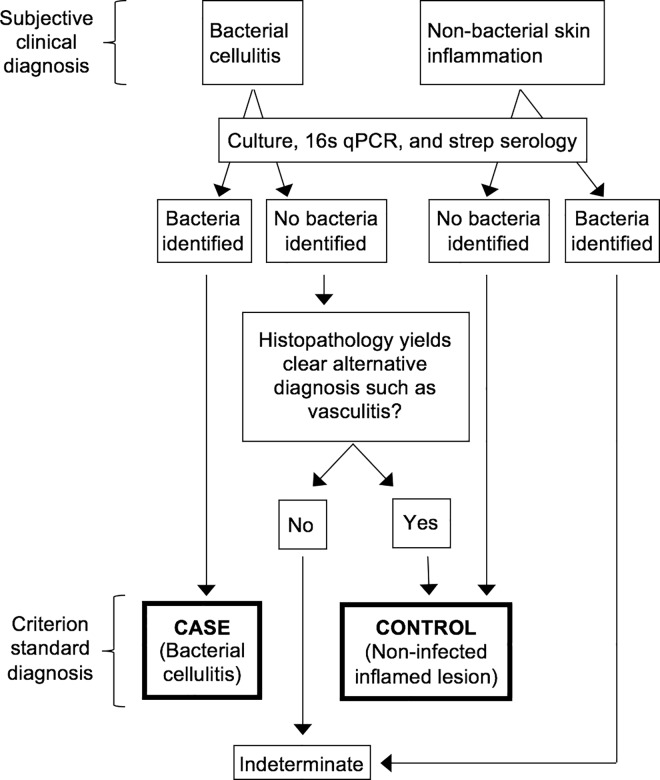
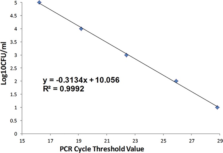
Methods: We classified lesions via biopsies, 16s rRNA gene detection, culture, and histopathology. We quantified procalcitonin expression in blood. We also used Nanostring technology to quantify transcription of immunomodulators that may distinguish cases from inflamed controls.

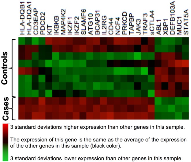
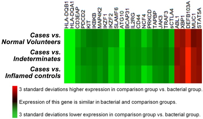
Results: Of 28 participants, 15 had a clinical diagnosis of cellulitis, six had a diagnosis of non-infectious dermatitis, and seven were normal volunteers. Of the "cellulitis" patients, three (20%) had pathogens isolated, and were designated confirmed cases. Procalcitonin was undetectable in all three. HLA-DQA1 was expressed 34-fold more in confirmed cases vs. controls (fold change of geometric mean). Heat maps depicting multiplex gene expression analysis revealed a distinct profile of gene expression in confirmed cases relative to comparators.

Conclusions: Most "cellulitis" patients had microbiologically-negative biopsies. Procalcitonin was undetectable, and HLA-DQA1 elevated, in confirmed bacterial cases. Multivariable transcriptomic profiling results supported our algorithm's ability to identify patients with true bacterial cellulitis. A larger sample may allow discovery of an immunological signature capable of distinguishing bacterial cellulitis from its mimics in clinical practice.


Conflict of interest statement

The authors received funding for this study from Biomerieux Inc., a commercial company and the manufacturer of procalcitonin assay utilized in the study. There are no other patents, products in development or marketed products to declare. This does not alter our adherence to all the PLOS ONE policies on sharing data and materials.

Figures

Fig 1
Fig 1. Algorithm for Identification of Confirmed Cellulitis Cases and Controls.
Please see the text for an explanation of the application of this algorithm.
Fig 2
Fig 2. Assessment of qPCR Sensitivity for Bacteria Using S. epidermdis.
We assessed the sensitivity of quantitative PCR for detection of bacteria via amplification of the universal bacterial 16s ribosomal RNA gene, using DNA purified from germ free mice skin tissue spiked with serial 10-fold dilutions of quantified Staphylococcus epidermidis. The assay's sensitivity was at or better than 10 colony-forming units per mL, with no evidence of decreasing sensitivity at lower levels.
Fig 3
Fig 3. Heat Map of a Derivation Set, Comprised of Verified Cases and Inflamed Controls.
This heat map, built on agglomerative clustering with linkage by mean and a Euclidian distance metric, shows a comparison of log(2)-transformed normalized gene expression data from blood. Rows represent individual genes, the symbols of which are shown at right. Columns represent individual participants in our study. Colors represent the z-scores of the counts of mRNA molecules per 100 ng RNA in blood; red represents a high ratio of expression relative to other genes in that participant, and green represents a low ratio of expression relative to other genes in that participant. The left three columns are confirmed bacterial cellulitis cases, and have high expression of genes HLA-DQB1 through sCTLA4, and low expression of genes ABL1 through STAT5A. All of the other columns are inflamed controls, as defined in the text.
Fig 4
Fig 4. Comparison of Gene Expression Patterns in Verified Bacterial Cases versus Three Comparison Groups.
This heat map, built on agglomerative clustering with linkage by mean and a Euclidian distance metric, shows a comparison of log(2)-transformed normalized gene expression data from blood. Rows represent individual genes, the symbols of which are shown at right. Each column displays a comparison of gene expression in bacterial cases (B) vs. one of three comparator groups: sterile inflamed controls (ic), indeterminate participants (ind), and normal volunteers (v). Qualitatively, this heat map reveals that the cases differ in a similar way from all three comparison groups. This, in turn, suggests that the cases–which had confirmed bacterial cellulitis based on biopsy–could be detectable by transcription profiling.
Fig 5
Fig 5. Photomicrographs from patients diagnosed clinically as having bacterial cellulitis.
Fig 5A: Culture-proven MRSA cellulitis with positive Gram stain. Note intracellular Gram-positive cocci in clusters. Fig 5B: Lymphocytic vasculitis misdiagnosed as cellulitis. Note abundant lymphocytes invading vessel wall.

References

    1. Chambers HF (2013) Cellulitis, by any other name. Clinical infectious diseases: an official publication of the Infectious Diseases Society of America 56: 1763–1764. - PubMed
    1. Arakaki RY, Strazzula L, Woo E, Kroshinsky D (2014) The impact of dermatology consultation on diagnostic accuracy and antibiotic use among patients with suspected cellulitis seen at outpatient internal medicine offices: a randomized clinical trial. JAMA dermatology 150: 1056–1061. 10.1001/jamadermatol.2014.1085 - DOI - PubMed
    1. David CV, Chira S, Eells SJ, Ladrigan M, Papier A, et al. (2011) Diagnostic accuracy in patients admitted to hospitals with cellulitis. Dermatol Online J 17: 1. - PubMed
    1. Strazzula L, Cotliar J, Fox LP, Hughey L, Shinkai K, et al. (2015) Inpatient dermatology consultation aids diagnosis of cellulitis among hospitalized patients: A multi-institutional analysis. Journal of the American Academy of Dermatology 73: 70–75. 10.1016/j.jaad.2014.11.012 - DOI - PubMed
    1. Pallin DJ, Camargo CA Jr., Schuur JD (2014) Skin infections and antibiotic stewardship: analysis of emergency department prescribing practices, 2007–2010. The western journal of emergency medicine 15: 282–289. 10.5811/westjem.2013.8.18040 - DOI - PMC - PubMed