Review
. 2016 Sep 20;8(9):257.
doi: 10.3390/v8090257.

Orchestrating the Selection and Packaging of Genomic RNA by Retroviruses: An Ensemble of Viral and Host Factors

Affiliations
Review

Orchestrating the Selection and Packaging of Genomic RNA by Retroviruses: An Ensemble of Viral and Host Factors

Rebecca J Kaddis Maldonado et al. Viruses. .

Abstract

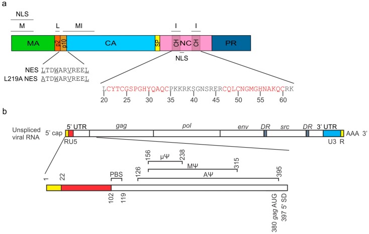
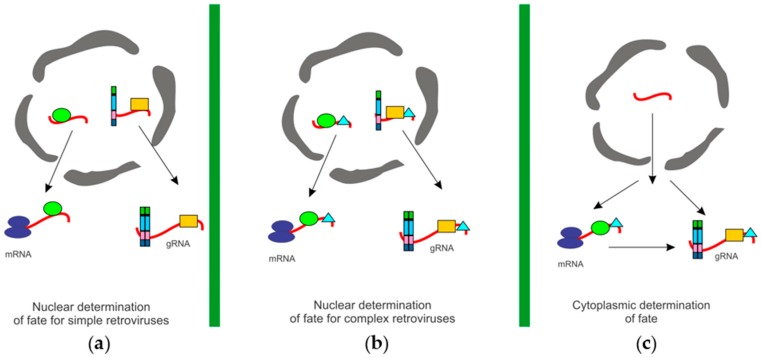
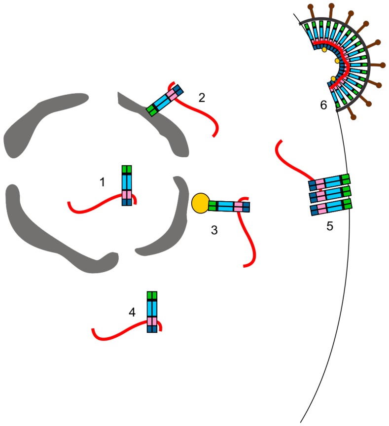
Infectious retrovirus particles contain two copies of unspliced viral RNA that serve as the viral genome. Unspliced retroviral RNA is transcribed in the nucleus by the host RNA polymerase II and has three potential fates: (1) it can be spliced into subgenomic messenger RNAs (mRNAs) for the translation of viral proteins; or it can remain unspliced to serve as either (2) the mRNA for the translation of Gag and Gag-Pol; or (3) the genomic RNA (gRNA) that is packaged into virions. The Gag structural protein recognizes and binds the unspliced viral RNA to select it as a genome, which is selected in preference to spliced viral RNAs and cellular RNAs. In this review, we summarize the current state of understanding about how retroviral packaging is orchestrated within the cell and explore potential new mechanisms based on recent discoveries in the field. We discuss the cis-acting elements in the unspliced viral RNA and the properties of the Gag protein that are required for their interaction. In addition, we discuss the role of host factors in influencing the fate of the newly transcribed viral RNA, current models for how retroviruses distinguish unspliced viral mRNA from viral genomic RNA, and the possible subcellular sites of genomic RNA dimerization and selection by Gag. Although this review centers primarily on the wealth of data available for the alpharetrovirus Rous sarcoma virus, in which a discrete RNA packaging sequence has been identified, we have also summarized the cis- and trans-acting factors as well as the mechanisms governing gRNA packaging of other retroviruses for comparison.

Keywords: retroviral Gag proteins; retrovirus assembly; retroviruses; subcellular trafficking; viral RNA export; viral RNA packaging; virus–cell interactions.


Conflict of interest statement

The authors declare no conflict of interest.

Figures

Figure 1
Figure 1
The Rous sarcoma virus (RSV) Gag polyprotein domain organization and functional sequences in the 5′ untranslated region (UTR) of the genomic RNA. (a) The RSV Gag protein is made of multiple domains: matrix (MA; green) contains the membrane binding motif (M) and a non-canonical nuclear localization signal (NLS); p2 (orange) contains the late (L) motif. p10 (gold) contains a chromosome region maintenance 1 (CRM1)-dependent nuclear export signal (NES); capsid (CA; blue) contains a multimerization interface (MI) that extends from p10; spacer peptide (SP; yellow); nucleocapsid (NC; pink) binds nucleic acids and proteins via interaction motifs (I) and Cys-His boxes (CH; translucent grey) and also contains an NLS; and protease (PR; blue). The Cys-His boxes contain zinc finger domains (red) required for the binding of the Ψ packaging sequence located in the 5′ UTR of the viral RNA (vRNA); (b) The primer binding site (PBS) is required for reverse transcription (nts 102–119). The entire RSV packaging sequence AΨ extends from nucleotides 126–395. The MΨ sequence extends from nucleotides 156–215. The minimal functional packaging element, identified as µΨ, is 82 nucleotides in length, extending from 156 to 238. Because the splice donor (SD) is located downstream of the packaging sequence at nucleotide 397, the Ψ sequence is contained in both spliced and unspliced vRNAs. DR: direct repeat; RU5: repeat-unique 5’; U3: unique 3’.
Figure 2
Figure 2
Models for unspliced retroviral RNA utilization. (a) Nuclear determination of vRNA fate for simple retroviruses: The cytoplasmic utilization of unspliced vRNAs may be determined by the co-transcriptional binding of specific factors that “mark” the unspliced vRNA for either packaging or translation, and the vRNAs are not interchangeable. The mechanism by which the unspliced RNA is exported from the nucleus could determine the cytoplasmic utilization of the unspliced retroviral RNA. Models include: (i) Nuclear Gag binds to the vRNA to mark it for packaging; (ii) Nuclear Gag and a host factor bind to the vRNA to mark it for packaging; or (iii) Nuclear host factors bind to the unspliced vRNA to determine its fate; (b) Nuclear determination of fate for complex retroviruses: Complex retroviruses encode accessory proteins that facilitate the nuclear export of unspliced or incompletely spliced retroviral RNA, such as HIV-1 Rev. In addition to Rev-like proteins, other host and viral factors also bind to the unspliced vRNA co-transcriptionally and may contribute to the fate of unspliced vRNA. Models include: (i) Nuclear viral export factor and Gag bind to the vRNA to mark it as a genome for packaging; (ii) Nuclear viral export factor, Gag, and a host factor bind to the vRNA to sort it for packaging; or (iii) Nuclear viral export factor and specific host factors bind to the unspliced vRNA to determine its cytoplasmic fate; (c) Cytoplasmic determination of fate: The fate of unspliced vRNAs may be defined in the cytoplasm. An unspliced vRNA can be used for either translation or packaging. A previously translated RNA can subsequently be packaged. In this scenario, host and viral factors may be bound to the unspliced vRNA in the nucleus, but their presence does not determine cytoplasmic utilization of the vRNA.
Figure 3
Figure 3
Potential subcellular sites of the initial Gag–gRNA interaction. Gag and the vRNA may first interact: (1) in the nucleus; (2) along the cytoplasmic face of the nuclear envelope; (3) at the microtubule organizing center (MTOC; yellow); (4) in the cytoplasm; or (5) at the plasma membrane. In each case, the viral ribonucleoprotein complex binds to the plasma membrane and interacts with other Gag proteins to form a virion that (6) buds from the plasma membrane.

Similar articles

Cited by

References

    1. Giorgi C., Moore M.J. The nuclear nurture and cytoplasmic nature of localized mRNPs. Semin. Cell Dev. Biol. 2007;18:186–193. doi: 10.1016/j.semcdb.2007.01.002. - DOI - PubMed
    1. Reed R. Coupling transcription, splicing and mRNA export. Curr. Opin. Cell Biol. 2003;15:326–331. doi: 10.1016/S0955-0674(03)00048-6. - DOI - PubMed
    1. Trcek T., Singer R.H. The cytoplasmic fate of an mRNP is determined cotranscriptionally: Exception or rule? Genes Dev. 2010;24:1827–1831. doi: 10.1101/gad.1972810. - DOI - PMC - PubMed
    1. Stoltzfus C.M. Synthesis and processing of avian sarcoma retrovirus RNA. Adv. Virus Res. 1988;35:1–38. - PubMed
    1. Coffin J.M., Hughes S.H., Varmus H.E. The interactions of retroviruses and their hosts. In: Coffin J.M., Hughes S.H., Varmus H.E., editors. Retroviruses. Cold Spring Harbor; New York, NY, USA: 1997. - PubMed

LinkOut - more resources