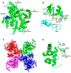
DNA Polymerase θ: A Unique Multifunctional End-Joining Machine
- PMID: 27657134
- PMCID: PMC5042397
- DOI: 10.3390/genes7090067
DNA Polymerase θ: A Unique Multifunctional End-Joining Machine
Abstract
The gene encoding DNA polymerase θ (Polθ) was discovered over ten years ago as having a role in suppressing genome instability in mammalian cells. Studies have now clearly documented an essential function for this unique A-family polymerase in the double-strand break (DSB) repair pathway alternative end-joining (alt-EJ), also known as microhomology-mediated end-joining (MMEJ), in metazoans. Biochemical and cellular studies show that Polθ exhibits a unique ability to perform alt-EJ and during this process the polymerase generates insertion mutations due to its robust terminal transferase activity which involves template-dependent and independent modes of DNA synthesis. Intriguingly, the POLQ gene also encodes for a conserved superfamily 2 Hel308-type ATP-dependent helicase domain which likely assists in alt-EJ and was reported to suppress homologous recombination (HR) via its anti-recombinase activity. Here, we review our current knowledge of Polθ-mediated end-joining, the specific activities of the polymerase and helicase domains, and put into perspective how this multifunctional enzyme promotes alt-EJ repair of DSBs formed during S and G2 cell cycle phases.
Keywords: DNA polymerase; DNA repair; alternative end-joining; cancer; genome instability; microhomology-mediated end-joining; replication repair.
Conflict of interest statement
R.T.P. has filed provisional patents on the use of Polθ for modifying the 3’ ends of nucleic acids and expanded-size nucleotide analogs as Polθ inhibitors and cancer therapeutics.
Figures


Similar articles
-
Structural basis for Polθ-helicase DNA binding and microhomology-mediated end-joining.Nat Commun. 2025 Apr 19;16(1):3725. doi: 10.1038/s41467-025-58441-x. Nat Commun. 2025. PMID: 40253368 Free PMC article.
-
Structural Basis for Polθ-Helicase DNA Binding and Microhomology-Mediated End-Joining.bioRxiv [Preprint]. 2024 Jun 7:2024.06.07.597860. doi: 10.1101/2024.06.07.597860. bioRxiv. 2024. Update in: Nat Commun. 2025 Apr 19;16(1):3725. doi: 10.1038/s41467-025-58441-x. PMID: 38895274 Free PMC article. Updated. Preprint.
-
Talazoparib enhances resection at DSBs and renders HR-proficient cancer cells susceptible to Polθ inhibition.Radiother Oncol. 2024 Nov;200:110475. doi: 10.1016/j.radonc.2024.110475. Epub 2024 Aug 13. Radiother Oncol. 2024. PMID: 39147034
-
DNA polymerase θ (POLQ), double-strand break repair, and cancer.DNA Repair (Amst). 2016 Aug;44:22-32. doi: 10.1016/j.dnarep.2016.05.003. Epub 2016 May 14. DNA Repair (Amst). 2016. PMID: 27264557 Free PMC article. Review.
-
Targeting Polymerase Theta (POLθ) for Cancer Therapy.Cancer Treat Res. 2023;186:285-298. doi: 10.1007/978-3-031-30065-3_15. Cancer Treat Res. 2023. PMID: 37978141 Review.
Cited by
-
RAD52 as a Potential Target for Synthetic Lethality-Based Anticancer Therapies.Cancers (Basel). 2019 Oct 14;11(10):1561. doi: 10.3390/cancers11101561. Cancers (Basel). 2019. PMID: 31615159 Free PMC article. Review.
-
Biomarker-Guided Development of DNA Repair Inhibitors.Mol Cell. 2020 Jun 18;78(6):1070-1085. doi: 10.1016/j.molcel.2020.04.035. Epub 2020 May 26. Mol Cell. 2020. PMID: 32459988 Free PMC article. Review.
-
Enzymatic synthesis of random sequences of RNA and RNA analogues by DNA polymerase theta mutants for the generation of aptamer libraries.Nucleic Acids Res. 2018 Jul 6;46(12):6271-6284. doi: 10.1093/nar/gky413. Nucleic Acids Res. 2018. PMID: 29788485 Free PMC article.
-
Genetic factors governing bacterial virulence and host plant susceptibility during Agrobacterium infection.Adv Genet. 2022;110:1-29. doi: 10.1016/bs.adgen.2022.08.001. Epub 2022 Sep 13. Adv Genet. 2022. PMID: 37283660 Free PMC article. Review.
-
Hybrid sequencing resolves two germline ultra-complex chromosomal rearrangements consisting of 137 breakpoint junctions in a single carrier.Hum Genet. 2021 May;140(5):775-790. doi: 10.1007/s00439-020-02242-3. Epub 2020 Dec 14. Hum Genet. 2021. PMID: 33315133 Free PMC article.
References
-
- Goff J.P., Shields D.S., Seki M., Choi S., Epperly M.W., Dixon T., Wang H., Bakkenist C.J., Dertinger S.D., Torous D.K., et al. Lack of DNA polymerase theta (POLQ) radiosensitizes bone marrow stromal cells in vitro and increases reticulocyte micronuclei after total-body irradiation. Radiat. Res. 2009;172:165–174. doi: 10.1667/RR1598.1. - DOI - PMC - PubMed
-
- Higgins G.S., Prevo R., Lee Y.F., Helleday T., Muschel R.J., Taylor S., Yoshimura M., Hickson I.D., Bernhard E.J., McKenna W.G. A small interfering RNA screen of genes involved in DNA repair identifies tumor-specific radiosensitization by POLQ knockdown. Cancer Res. 2010;70:2984–2993. doi: 10.1158/0008-5472.CAN-09-4040. - DOI - PMC - PubMed
Publication types
Grants and funding
LinkOut - more resources
Full Text Sources
Other Literature Sources

