

Cerenkov Radiation Induced Photodynamic Therapy Using Chlorin e6-Loaded Hollow Mesoporous Silica Nanoparticles
- PMID: 27657487
- PMCID: PMC5061626
- DOI: 10.1021/acsami.6b10255
Cerenkov Radiation Induced Photodynamic Therapy Using Chlorin e6-Loaded Hollow Mesoporous Silica Nanoparticles
Abstract
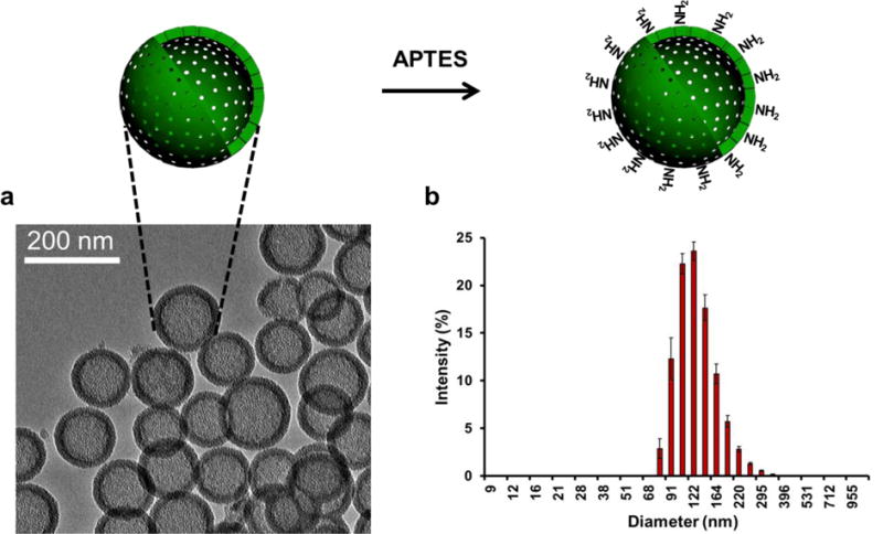
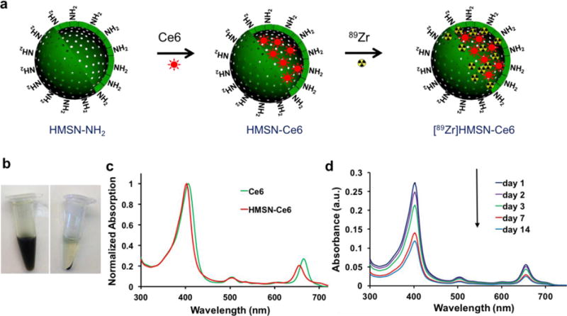
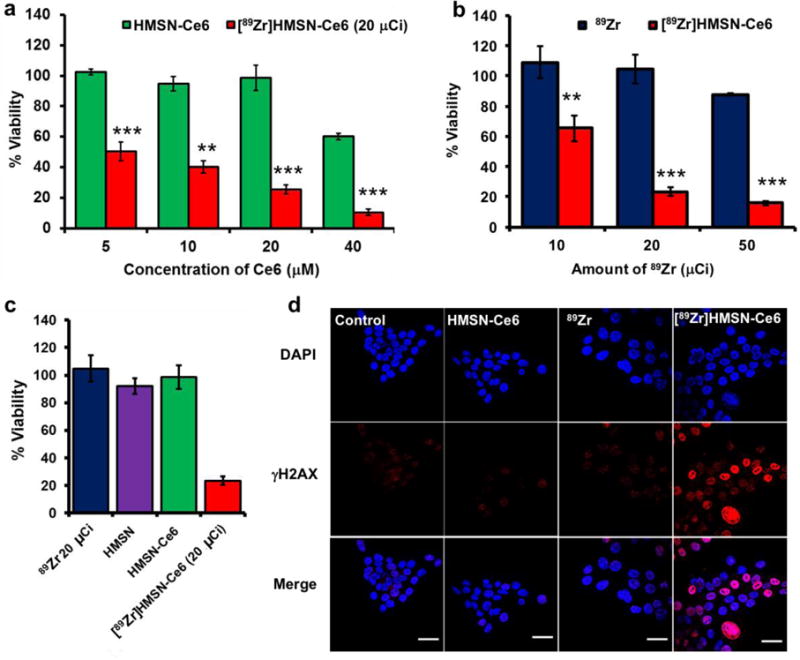
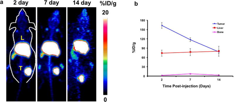
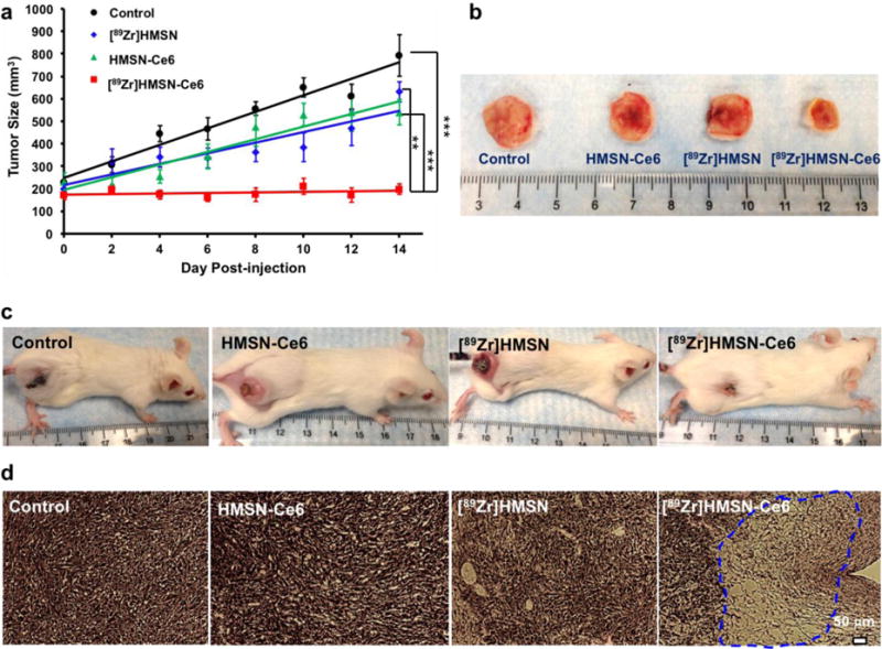
Traditional photodynamic therapy (PDT) requires external light to activate photosensitizers for therapeutic purposes. However, the limited tissue penetration of light is still a major challenge for this method. To overcome this limitation, we report an optimized system that uses Cerenkov radiation for PDT by using radionuclides to activate a well-known photosensitizer (chlorin e6, Ce6). By taking advantage of hollow mesoporous silica nanoparticles (HMSNs) that can intrinsically radiolabel an oxophilic zirconium-89 (89Zr, t1/2 = 78.4 h) radionuclide, as well as possess great drug loading capacity, Ce6 can be activated by Cerenkov radiation from 89Zr in the same nanoconstruct. In vitro cell viability experiments demonstrated dose-dependent cell deconstruction as a function of the concentration of Ce6 and 89Zr. In vivo studies show inhibition of tumor growth when mice were subcutaneously injected with [89Zr]HMSN-Ce6, and histological analysis of the tumor section showed damage to tumor tissues, implying that reactive oxygen species mediated the destruction. This study offers a way to use an internal radiation source to achieve deep-seated tumor therapy without using any external light source for future applications.
Keywords: Cerenkov radiation; chlorin e6; hollow mesoporous silica nanoparticles; photodynamic therapy; positron emission tomography.
Figures
Similar articles
-
Chlorin e6-loaded goat milk-derived extracellular vesicles for Cerenkov luminescence-induced photodynamic therapy.Eur J Nucl Med Mol Imaging. 2023 Jan;50(2):508-524. doi: 10.1007/s00259-022-05978-4. Epub 2022 Oct 12. Eur J Nucl Med Mol Imaging. 2023. PMID: 36222853
-
Titania and silica nanoparticles coupled to Chlorin e6 for anti-cancer photodynamic therapy.Photodiagnosis Photodyn Ther. 2018 Jun;22:115-126. doi: 10.1016/j.pdpdt.2018.03.005. Epub 2018 Mar 23. Photodiagnosis Photodyn Ther. 2018. PMID: 29581041
-
Co-Delivery of Cisplatin Prodrug and Chlorin e6 by Mesoporous Silica Nanoparticles for Chemo-Photodynamic Combination Therapy to Combat Drug Resistance.ACS Appl Mater Interfaces. 2016 Jun 1;8(21):13332-40. doi: 10.1021/acsami.6b03881. Epub 2016 May 17. ACS Appl Mater Interfaces. 2016. PMID: 27164222
-
Scintillating Nanoparticles as Energy Mediators for Enhanced Photodynamic Therapy.ACS Nano. 2016 Apr 26;10(4):3918-35. doi: 10.1021/acsnano.6b01401. Epub 2016 Apr 8. ACS Nano. 2016. PMID: 27043181 Free PMC article. Review.
-
Radiolabeling Silica-Based Nanoparticles via Coordination Chemistry: Basic Principles, Strategies, and Applications.Acc Chem Res. 2018 Mar 20;51(3):778-788. doi: 10.1021/acs.accounts.7b00635. Epub 2018 Feb 28. Acc Chem Res. 2018. PMID: 29489335 Free PMC article. Review.
Cited by
-
Amplification of Cerenkov luminescence using semiconducting polymers for cancer theranostics.Adv Funct Mater. 2023 Aug 15;33(33):2302777. doi: 10.1002/adfm.202302777. Epub 2023 May 1. Adv Funct Mater. 2023. PMID: 37942189 Free PMC article.
-
A "Missile-Detonation" Strategy to Precisely Supply and Efficiently Amplify Cerenkov Radiation Energy for Cancer Theranostics.Adv Mater. 2019 Dec;31(52):e1904894. doi: 10.1002/adma.201904894. Epub 2019 Nov 11. Adv Mater. 2019. PMID: 31709622 Free PMC article.
-
Radiation-Activated Cobalamin-Kinase Inhibitors for Treatment of Pancreatic Ductal Adenocarcinoma.Mol Pharm. 2024 Jan 1;21(1):137-142. doi: 10.1021/acs.molpharmaceut.3c00667. Epub 2023 Nov 21. Mol Pharm. 2024. PMID: 37989273 Free PMC article.
-
Minimizing adverse effects of Cerenkov radiation induced photodynamic therapy with transformable photosensitizer-loaded nanovesicles.J Nanobiotechnology. 2022 Apr 27;20(1):203. doi: 10.1186/s12951-022-01401-0. J Nanobiotechnology. 2022. PMID: 35477389 Free PMC article.
-
Activatable Hybrid Nanotheranostics for Tetramodal Imaging and Synergistic Photothermal/Photodynamic Therapy.Adv Mater. 2018 Feb;30(6):10.1002/adma.201704367. doi: 10.1002/adma.201704367. Epub 2017 Dec 21. Adv Mater. 2018. PMID: 29266476 Free PMC article.
References
-
- Triesscheijn M, Baas P, Schellens JH, Stewart FA. Photodynamic Therapy in Oncology. Oncologist. 2006;11:1034–1044. - PubMed
-
- Wani S, Puli SR, Shaheen NJ, Westhoff B, Slehria S, Bansal A, Rastogi A, Sayana H, Sharma P. Esophageal Adenocarcinoma in Barrett’s Esophagus after Endoscopic Ablative Therapy: A Meta-Analysis Systematic Review. Am J Gastroenterol. 2009;104:502–513. - PubMed
-
- Hsu CY, Chen CW, Yu HP, Lin YF, Lai PS. Bioluminescence Resonance Energy Transfer Using Luciferase-Immobilized Quantum Dots for Self-Illuminated Photodynamic Therapy. Biomaterials. 2013;34:1204–1212. - PubMed
-
- Magalhães CM, Esteves da Silva JCG, Pinto da Silva L. Chemiluminescence and Bioluminescence as an Excitation Source in the Photodynamic Therapy of Cancer: A Critical Review. ChemPhysChem. 2016;17:2286–2294. - PubMed
MeSH terms
Substances
Grants and funding
LinkOut - more resources
Full Text Sources
Other Literature Sources
