

Recombinant Atrial Natriuretic Peptide Prevents Aberrant Ca2+ Leakage through the Ryanodine Receptor by Suppressing Mitochondrial Reactive Oxygen Species Production Induced by Isoproterenol in Failing Cardiomyocytes
- PMID: 27657534
- PMCID: PMC5033569
- DOI: 10.1371/journal.pone.0163250
Recombinant Atrial Natriuretic Peptide Prevents Aberrant Ca2+ Leakage through the Ryanodine Receptor by Suppressing Mitochondrial Reactive Oxygen Species Production Induced by Isoproterenol in Failing Cardiomyocytes
Abstract
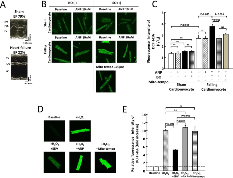
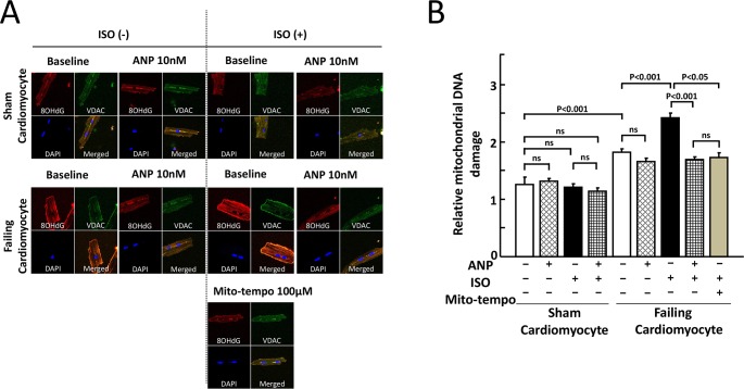
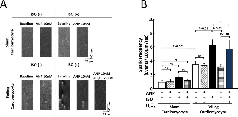
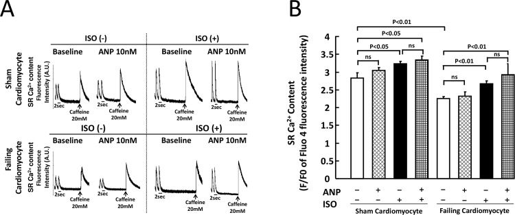
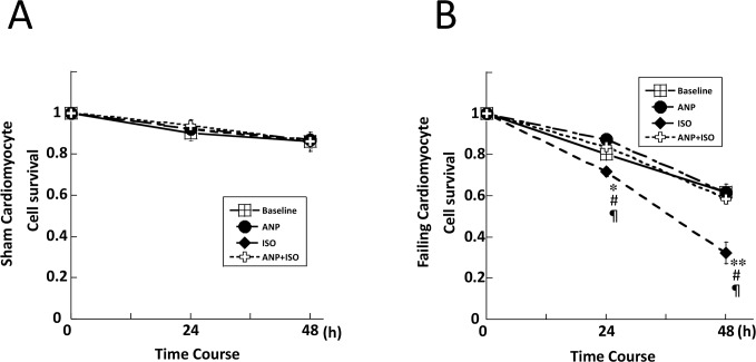
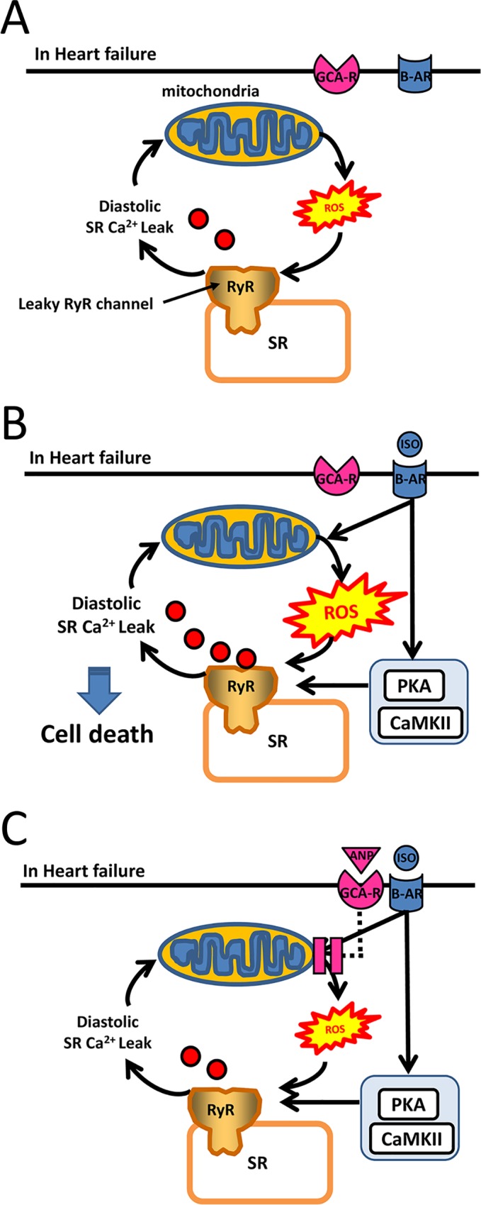
Catecholamines induce intracellular reactive oxygen species (ROS), thus enhancing diastolic Ca2+ leakage through the ryanodine receptor during heart failure (HF). However, little is known regarding the effect of atrial natriuretic peptide (ANP) on ROS generation and Ca2+ handling in failing cardiomyocytes. The aim of the present study was to clarify the mechanism by which an exogenous ANP exerts cardioprotective effects during HF. Cardiomyocytes were isolated from the left ventricles of a canine tachycardia-induced HF model and sham-operated vehicle controls. The degree of mitochondrial oxidized DNA was evaluated by double immunohistochemical (IHC) staining using an anti-VDAC antibody for the mitochondria and an anti-8-hydroxy-2'-deoxyguanosine antibody for oxidized DNA. The effect of ANP on ROS was investigated using 2,7-dichlorofluorescin diacetate, diastolic Ca2+ sparks assessed by confocal microscopy using Fluo 4-AM, and the survival rate of myocytes after 48 h. The double IHC study revealed that isoproterenol (ISO) markedly increased oxidized DNA in the mitochondria in HF and that the ISO-induced DNA damage was markedly inhibited by the co-presence of ANP. ROS production and Ca2+ spark frequency (CaSF) were increased in HF compared to normal controls, and were further increased in the presence of ISO. Notably, ANP significantly suppressed both ISO-induced ROS and CaSF without changing sarcoplasmic reticulum Ca2+ content in HF (p<0.01, respectively). The survival rate after 48 h in HF was significantly decreased in the presence of ISO compared with baseline (p<0.01), whereas it was significantly improved by the co-presence of ANP (p<0.01). Together, our results suggest that ANP strongly suppresses ISO-induced mitochondrial ROS generation, which might correct aberrant diastolic Ca2+ sparks, eventually contributing to the improvement of cardiomyocyte survival in HF.
Conflict of interest statement
The authors have declared that no competing interests exist.
Figures
Similar articles
-
Reconciling depressed Ca2+ sparks occurrence with enhanced RyR2 activity in failing mice cardiomyocytes.J Gen Physiol. 2015 Oct;146(4):295-306. doi: 10.1085/jgp.201511366. Epub 2015 Sep 14. J Gen Physiol. 2015. PMID: 26371209 Free PMC article.
-
Bidirectional regulation of Ca2+ sparks by mitochondria-derived reactive oxygen species in cardiac myocytes.Cardiovasc Res. 2008 Jan 15;77(2):432-41. doi: 10.1093/cvr/cvm047. Epub 2007 Oct 25. Cardiovasc Res. 2008. PMID: 18006452
-
Phosphodiesterase-2 is up-regulated in human failing hearts and blunts β-adrenergic responses in cardiomyocytes.J Am Coll Cardiol. 2013 Oct 22;62(17):1596-606. doi: 10.1016/j.jacc.2013.05.057. Epub 2013 Jun 26. J Am Coll Cardiol. 2013. PMID: 23810893
-
Calcium sparks in human ventricular cardiomyocytes from patients with terminal heart failure.Cell Calcium. 2002 Apr;31(4):175-82. doi: 10.1054/ceca.2002.0272. Cell Calcium. 2002. PMID: 12027382
-
Navigating Calcium and Reactive Oxygen Species by Natural Flavones for the Treatment of Heart Failure.Front Pharmacol. 2021 Nov 9;12:718496. doi: 10.3389/fphar.2021.718496. eCollection 2021. Front Pharmacol. 2021. PMID: 34858167 Free PMC article. Review.
Cited by
-
Empagliflozin suppressed cardiac fibrogenesis through sodium-hydrogen exchanger inhibition and modulation of the calcium homeostasis.Cardiovasc Diabetol. 2023 Feb 6;22(1):27. doi: 10.1186/s12933-023-01756-0. Cardiovasc Diabetol. 2023. PMID: 36747205 Free PMC article.
-
Regulation of mitochondria function by natriuretic peptides.Am J Physiol Renal Physiol. 2019 Nov 1;317(5):F1164-F1168. doi: 10.1152/ajprenal.00384.2019. Epub 2019 Sep 11. Am J Physiol Renal Physiol. 2019. PMID: 31509010 Free PMC article. Review.
-
Effects of BNP and Sacubitrilat/Valsartan on Atrial Functional Reserve and Arrhythmogenesis in Human Myocardium.Front Cardiovasc Med. 2022 Jul 5;9:859014. doi: 10.3389/fcvm.2022.859014. eCollection 2022. Front Cardiovasc Med. 2022. PMID: 35865376 Free PMC article.
-
Enhanced oxidative stress and presence of ventricular aneurysm for risk prediction in cardiac sarcoidosis.Heart. 2022 Mar;108(6):429-437. doi: 10.1136/heartjnl-2021-320244. Epub 2022 Jan 25. Heart. 2022. PMID: 35078868 Free PMC article.
-
Acute isoproterenol leads to age-dependent arrhythmogenesis in guinea pigs.Am J Physiol Heart Circ Physiol. 2018 Oct 1;315(4):H1051-H1062. doi: 10.1152/ajpheart.00061.2018. Epub 2018 Jul 20. Am J Physiol Heart Circ Physiol. 2018. PMID: 30028197 Free PMC article.
References
-
- Mann DL, Kent RL, Parsons B, Cooper G 4th. Adrenergic effects on the biology of the adult mammalian cardiocyte. Circulation. 1992;85: 790–804. - PubMed
-
- Curran J, Hinton MJ, Ríos E, Bers DM, Shannon TR. Beta-adrenergic enhancement of sarcoplasmic reticulum calcium leak in cardiac myocytes is mediated by calcium/calmodulin-dependent protein kinase. Circ Res. 2007;100: 391–398. - PubMed
LinkOut - more resources
Full Text Sources
Other Literature Sources
Research Materials
Miscellaneous
