

Analysis of Discrepancy Between Diagnostic Clinical Examination Findings and Corresponding Evaluation of Digital Images in the Telemedicine Approaches to Evaluating Acute-Phase Retinopathy of Prematurity Study
- PMID: 27657673
- PMCID: PMC5989319
- DOI: 10.1001/jamaophthalmol.2016.3502
Analysis of Discrepancy Between Diagnostic Clinical Examination Findings and Corresponding Evaluation of Digital Images in the Telemedicine Approaches to Evaluating Acute-Phase Retinopathy of Prematurity Study
Abstract
Importance: As effective treatments for potentially blinding retinopathy of prematurity (ROP) have been introduced, the importance of consistency in findings has increased, especially with the shift toward retinal imaging in infants at risk of ROP.
Objective: To characterize discrepancies in findings of ROP between digital retinal image grading and examination results from the Telemedicine Approaches to Evaluating Acute-Phase Retinopathy of Prematurity study, conducted from May 2011 to October 2013.
Design, setting, and participants: A poststudy consensus review of images was conducted by 4 experts, who examined discrepancies in findings between image grades by trained nonphysician readers and physician examination results in infants with referral-warranted ROP (RW-ROP). Images were obtained from 13 North American neonatal intensive care units from eyes of infants with birth weights less than 1251 g. For discrepancy categories with more than 100 cases, 40 were randomly selected; in total, 188 image sets were reviewed.
Main outcomes and measures: Consensus evaluation of discrepant image and examination findings for RW-ROP components.
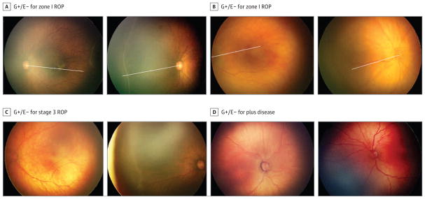
Results: Among 5350 image set pairs, there were 161 instances in which image grading did not detect RW-ROP noted on clinical examination (G-/E+) and 854 instances in which grading noted RW-ROP when the examination did not (G+/E-). Among the sample of G-/E+ cases, 18 of 32 reviews (56.3%) agreed with clinical examination findings that ROP was present in zone I and 18 of 40 (45.0%) agreed stage 3 ROP was present, but only 1 of 20 (5.0%) agreed plus disease was present. Among the sample of G+/E- cases, 36 of 40 reviews (90.0%) agreed with readers that zone I ROP was present, 23 of 40 (57.5%) agreed with readers that stage 3 ROP was present, and 4 of 16 (25.0%) agreed that plus disease was present. Based on the consensus review results of the sampled cases, we estimated that review would agree with clinical examination findings in 46.5% of the 161 G-/E+ cases (95% CI, 41.6-51.6) and agree with trained reader grading in 70.0% of the 854 G+/E- cases (95% CI, 67.3-72.8) for the presence of RW-ROP.
Conclusions and relevance: This report highlights limitations and strengths of both the remote evaluation of fundus images and bedside clinical examination of infants at risk for ROP. These findings highlight the need for standardized approaches as ROP telemedicine becomes more widespread.
Conflict of interest statement
Figures

Comment in
-
Telemedicine for Retinopathy of Prematurity: An Evolving Paradigm.JAMA Ophthalmol. 2016 Nov 1;134(11):1270-1271. doi: 10.1001/jamaophthalmol.2016.3518. JAMA Ophthalmol. 2016. PMID: 27657491 No abstract available.
References
-
- Cryotherapy for Retinopathy of Prematurity Cooperative Group. Multicenter trial of cryotherapy for retinopathy of prematurity: three-month outcome. Arch Ophthalmol. 1990;108(2):195–204. - PubMed
-
- Gschließer A, Stifter E, Neumayer T, et al. Inter-expert and intra-expert agreement on the diagnosis and treatment of retinopathy of prematurity. Am J Ophthalmol. 2015;160(3):553–560. e3. - PubMed
-
- Chiang MF, Wang L, Busuioc M, et al. Telemedical retinopathy of prematurity diagnosis: accuracy, reliability, and image quality. Arch Ophthalmol. 2007;125(11):1531–1538. - PubMed
Publication types
MeSH terms
Grants and funding
LinkOut - more resources
Full Text Sources
Other Literature Sources
Medical
Research Materials
