. 2016 Nov 1;134(11):1272-1280.
doi: 10.1001/jamaophthalmol.2016.3519.

Histopathological Insights Into Choroidal Vascular Loss in Clinically Documented Cases of Age-Related Macular Degeneration

Affiliations

Histopathological Insights Into Choroidal Vascular Loss in Clinically Documented Cases of Age-Related Macular Degeneration

Johanna M Seddon et al. JAMA Ophthalmol. .

Abstract

Importance: Age-related macular degeneration (AMD) is a multifactorial disease with genetic and environmental factors contributing to risk. Histopathologic changes underlying AMD are not fully understood, particularly the relationship between choriocapillaris (CC) dysfunction and phenotypic variability of this disease.

Objective: To examine histopathologic changes in the CC of eyes with clinically documented AMD.

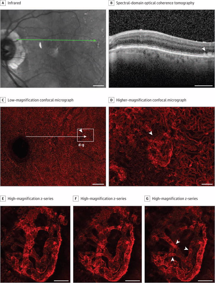
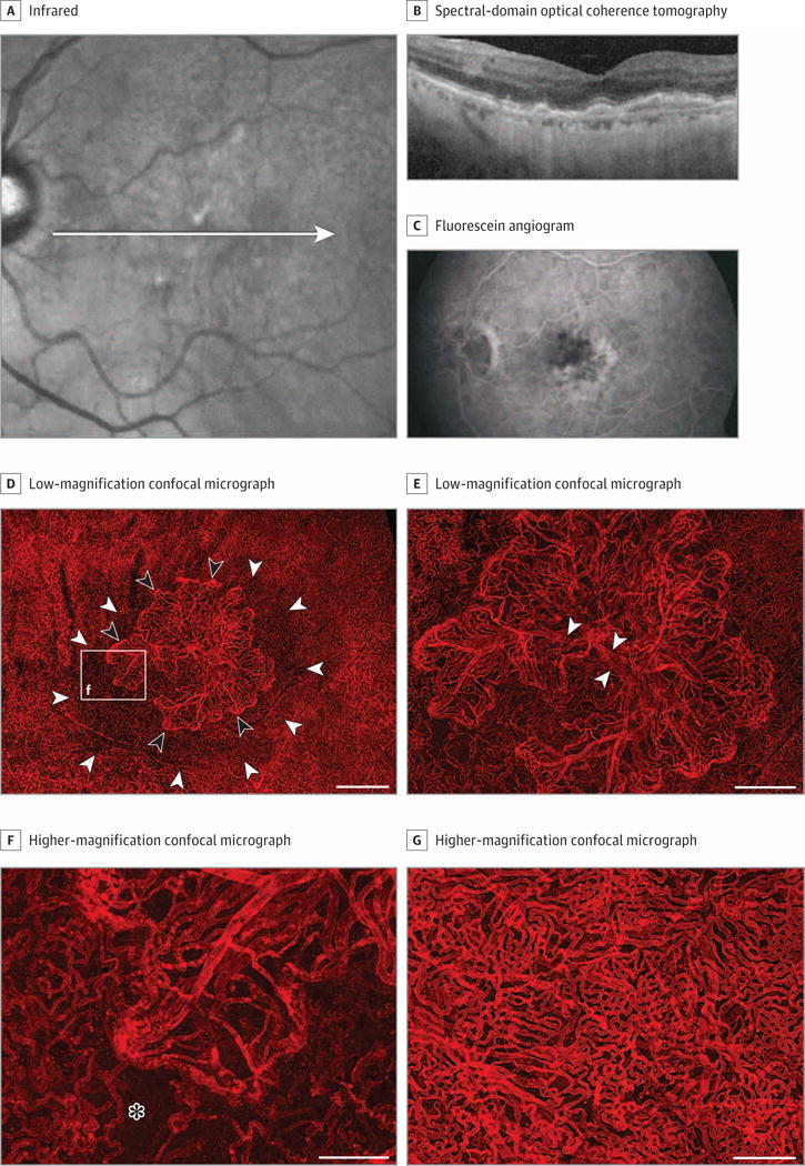
Design, setting, and participants: The study was designed in 2011. Tissues were collected post mortem (2012-2016), and histopathological images were obtained from participants enrolled in AMD studies since 1988. Clinical records and images were collected from participants as standard protocol. Eyes without AMD (n = 4) and eyes with early (n = 9), intermediate (n = 5), and advanced stages of AMD (geographic atrophy, n = 5; neovascular disease, n = 13) were evaluated. Choroidal vasculature was labeled using Ulex europaeus agglutinin lectin and examined using confocal microscopy.

Main outcomes and measures: A standardized classification system was applied to determine AMD stage. Ocular records and images were reviewed and histopathologic analyses performed. Viability of the choroidal vasculature was analyzed for each AMD stage.

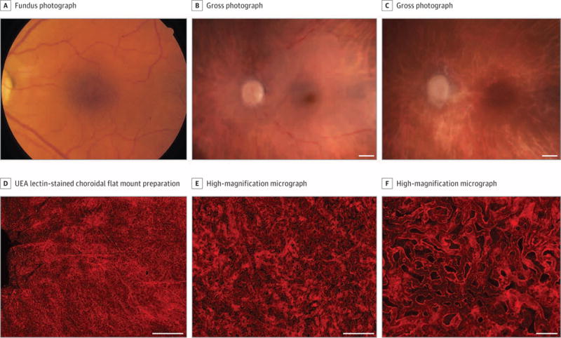
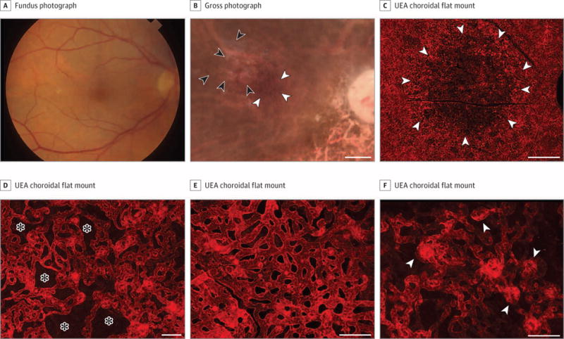
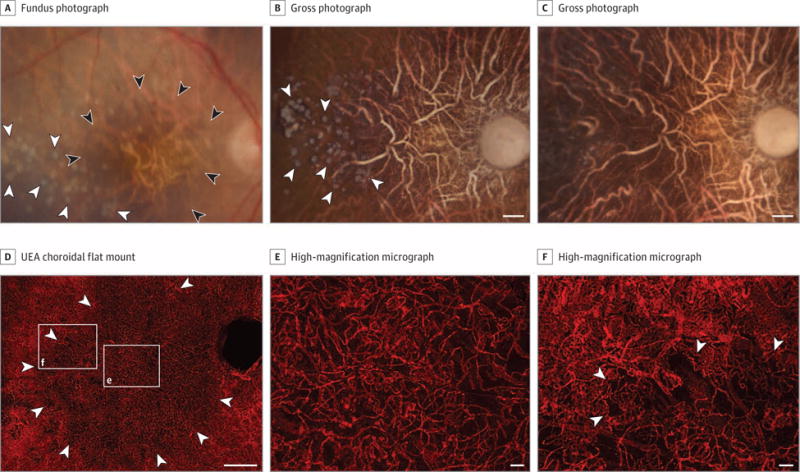
Results: All participants were white. Fourteen were male, and 16 were female. The mean age was 90.5 years among AMD patients and 88.5 years among control participants. Submacular CC dropout without retinal pigment eipthelial (RPE) loss was observed in all cases with early stages of AMD. Higher vascular area loss for each AMD stage was observed compared with control participants: 20.5% in early AMD (95% CI, 11.2%-40.2%; P < .001), 12.5% in intermediate AMD (95% CI, 2.9%-21.4%; P = .01), 39.0% loss in GA (95% CI, 32.1%-45.4%; P < .001), and 38.2% loss in neovascular disease where RPE remained intact (95% CI, 27.7%-47.9%; P < .001). Hypercellular, apparent neovascular buds were adjacent to areas of CC loss in 22.2% of eyes with early AMD and 40% of eyes with intermediate AMD.

Conclusions and relevance: Retinal pigment epithelial atrophy preceded CC loss in geographic atrophy, but CC loss occurred in the absence of RPE atrophy in 2 of 9 eyes with early-stage AMD. Given the cross-sectional nature of this study and the small number of eyes evaluated, definitive conclusions regarding this progression cannot be determined with certainty. We speculate that neovascular buds may be a precursor to neovascular disease. Hypoxic RPE resulting from reduced blood supply might upregulate production of vascular endothelial growth factor, providing the stimulus for neovascular disease.


Conflict of interest statement

Conflict of Interest Disclosures: All authors have completed and submitted the ICMJE Form for Disclosure of Potential Conflicts of Interest and none were reported.

Figures

Figure 1
Figure 1. Grade 1, Patient 3, Left Eye
Fundus photograph (A), gross photographs (B-C), and Ulex europaeus agglutinin (UEA) lectin-stained choroidal flat mount preparation (D-F) from an 87-year-old white woman with no history of age-related macular degeneration. The fundus photograph (A) shows a normal appearance of the macula. Postmortem gross photographs of the posterior eyecup with the retina intact (B) and the retina removed (C) show absence of drusen, retinal pigment epithelium loss, or hyperpigmentation. Low-magnification confocal micrograph of the UEA choroidal flat mount shows normal homogeneous pattern of choriocapillaris in the posterior pole region (D). Higher-magnification micrographs (E-F) show broad-diameter freely interconnecting capillaries. The mean (SD) percent vascular area in the submacula was 77.8% (3.5%) and the mean (SD) choriocapillaris diameter was 12.8 (1.2) μm. Scale bars: B-D = 1 mm, E = 200 μm; F = 50 μm.
Figure 2
Figure 2. Grade 2C, Patient 10, Right Eye
Fundus photograph (A), gross photograph (B), and Ulex europaeus agglutinin (UEA) lectin-stained choroidal flat mount preparation (C-F) from an 86-year-old white woman with a history of early-stage AMD. The fundus photograph (A) shows drusen in the macula. Postmortem gross photograph with the retina removed (B) shows presence of drusen (black arrowheads) and some pigmentary abnormalities (white arrowheads). Low-magnification confocal micrograph of the UEA choroidal flat mount shows a region of choriocapillaris (CC) pathology (arrowheads) in the submacula that was 10.5 mm2 (C). Higher-magnification micrographs show loss of interconnecting capillary channels (asterisks) and thinning of the CC lumen in submacula (D) compared with broad-diameter freely interconnecting capillaries in the perimacular region (E). At the border of submacular CC atrophy, small apparent neovascular buds (arrowheads) were observed (F). The mean (SD) percent vascular area was 43% (8.7%) in the submacula compared with 76% (2.8%) in the perimacular region. The mean (SD) CC diameter was 13.5 (0.7) μm in the submacular region compared to 17.4 (1.8) μm in the perimacular region. Scale bars: B and C = 1 mm, D-F = 50 μm.
Figure 3
Figure 3. Grade 3, Patient 13, Left Eye
Infrared image (A) and spectral-domain optical coherence tomography (OCT) (B) obtained 3 months prior to death, and Ulex europaeus agglutinin (UEA) lectin-stained choroidal flat mount preparation (C-G) from a 93-year-old white man with a history of intermediate AMD (grade 3). The infrared image shows numerous drusen in the posterior pole with some apparent pigmentary abnormalities (A) in the macula and large drusen (arrowhead) on OCT (B). Low-magnification confocal micrograph of the UEA choroidal flat mount (C) shows reduced choriocapillaris density throughout submacula. The long arrow is an approximation of the position of the green scan line in A. Higher-magnification micrograph (D) of the region indicated by box in C shows loss of interconnecting capillary channels and a vascular malformation (arrowhead) that corresponds to the region indicated by the arrow in the OCT. High-magnification Z-series (E-G) of the vascular formation shown in D demonstrates that it is internal to the choriocapillaris and represents a form of choroidal neovascularization. It has multiple connections to the underlying choroidal vasculature (arrowheads in G). Scale bars: A-C = 1 mm, D-F = 100 μm.
Figure 4
Figure 4. Grade 4, Patient 15, Right Eye
Fundus photograph (A), gross photographs (B and C), and Ulex europaeus agglutinin (UEA) lectin-stained choroidal flat mount preparation (D-F) from a 97-year-old white woman with a history of geographic atrophy. The fundus photograph (A) shows a well-defined area of retinal pigment epithelial (RPE) atrophy in the macula (black arrowheads) and drusen adjacent to the region of atrophy (white arrowheads). Postmortem gross photograph of the posterior eyecup with the retina removed prior to EDTA treatment (B) shows presence of glistening white calcified drusen in the RPE layer (arrowheads). Gross photograph of the posterior eyecup following EDTA treatment to remove RPE (C) shows loss of calcified drusen. Low-magnification confocal micrograph of the UEA choroidal flat mount shows region of choriocapillaris (CC) pathology (arrowheads) in the posterior pole region corresponding to area of RPE atrophy that was 37.5 mm2 (D). Higher-magnification micrographs show loss of interconnecting capillary channels and thinning of the CC lumen in the area of atrophy (box “e” in D) and at the border (arrowheads) of atrophy (box “f” in D). The mean (SD) percent vascular area was 38.3% (5%) in the area with RPE atrophy in the submacular region compared with 81.9% (23.9%) outside the area of RPE atrophy. The mean (SD) CC diameter was 9.3 (0.7) μm in the atrophic region compared with 19.5 (0.9) μm in the nonatrophic region. Scale bars: B-D = 1 mm, E and F = 50 μm.
Figure 5
Figure 5. Grade 5, Patient 23, Left Eye
Infrared and spectral-domain optical coherence tomography (OCT) images (A and B), a fluorescein angiogram (C), and Ulex europaeus agglutinin (UEA) lectin-stained choroid (D-G) from an 84-year-old white man with a history of neovascular disease treated with 6 anti–vascular endothelial growth factor injections. The infrared and OCT images (A and B) show drusen (including reticular-type drusen), retinal pigment epithelial (RPE) detachment, and early atrophic changes. The fluorescein angiogram prior to treatment displays the active choroidal neovascularization (CNV) (C). Low-magnification confocal micrograph of the UEA choroidal flat mount shows a CNV formation (black arrowheads) in the submacula, which was 5.8 mm2 (D and E). Choriocapillaris attenuation extended well beyond the edge of the CNV (white arrowheads in D). Higher-magnification micrograph (F) shows edge of CNV (area in box “f” in D) with looped hypercellular tips of neovascular vessels and capillary dropout adjacent to the edge of the CNV (asterisk). Peripheral to the CNV (G), the choriocapillaris has normal morphology. Scale bars: D = 1 mm, E = 500 μm, F and G = 250 μm.

Comment in

Similar articles

Cited by

References

    1. Friedman DS, O’Colmain BJ, Muñoz B, et al. Eye Diseases Prevalence Research Group Prevalence of age-related macular degeneration in the United States. Arch Ophthalmol. 2004;122(4):564–572. - PubMed
    1. Sobrin L, Seddon JM. Nature and nurture–genes and environment–predict onset and progression of macular degeneration. Prog Retin Eye Res. 2014;40:1–15. - PMC - PubMed
    1. Fritsche LG, Chen W, Schu M, et al. AMD Gene Consortium Seven new loci associated with age-related macular degeneration. Nat Genet. 2013;45(4):433–439. e1–e2. - PMC - PubMed
    1. Lim LS, Mitchell P, Seddon JM, Holz FG, Wong TY. Age-related macular degeneration. Lancet. 2012;379(9827):1728–1738. - PubMed
    1. McLeod DS, Taomoto M, Otsuji T, Green WR, Sunness JS, Lutty GA. Quantifying changes in RPE and choroidal vasculature in eyes with age-related macular degeneration. Invest Ophthalmol Vis Sci. 2002;43(6):1986–1993. - PubMed

MeSH terms